Contact/News Media

Wednesday, October 29, 2014

Teeth, sex and testosterone reveal secrets of aging in wild mouse lemurs

A brown mouse lemur in the wild. Mouse lemurs, weighing a mere 30 to 80 grams, are the world's smallest primates. Photos, above and below, by Jukka Jernvall.

By Carol Clark

Mouse lemurs can live at least eight years in the wild – twice as long as some previous estimates, a long-term longitudinal study finds.

PLOS ONE published the research on brown mouse lemurs (Microcebus rufus) led in Madagascar by biologist Sarah Zohdy, a post-doctoral fellow in Emory's Department of Environmental Sciences and the Rollins School of Public Health. Zohdy conducted the research while she was a doctoral student at the University of Helsinki.

“It’s surprising that these tiny, mouse-sized primates, living in a jungle full of predators that probably consider them a bite-sized snack, can live so long,” Zohdy says. “And we found individuals up to eight years of age in the wild with no physical symptoms of senescence like some captive mouse lemurs start getting by the age of four.”

It is likely that starvation, predation, disease and other environmental stressors reduce the observed rate of senescence in the wild, Zohdy notes, but a growing body of evidence also suggests that captive conditions may affect mental and physical function.

“We focused on wild mouse lemurs because we want to know what happens naturally when a primitive primate is exposed to all of the extrinsic and intrinsic mortality factors that shaped them as a species,” Zohdy says. “Comparing longevity data of captive and wild mouse lemurs may help us understand how the physiological and behavioral demands of different environments affect the aging process in other primates, including humans.”

The study determined ages of wild mouse lemurs in Madagascar’s Ranomafana National Park through a dental mold method that had not previously been used with small mammals. In addition to the high-resolution tooth-wear analysis for aging, fecal samples underwent hormone analysis.


The researchers found no difference between the longevity of male and female mouse lemurs, unlike most vertebrates where males tend to die first.

“And even more interestingly, we found no difference in testosterone levels between males and females,” Zohdy says. Mouse lemurs are female dominant, which may explain why their testosterone levels are on a par with males.

“While elevated male testosterone levels have been implicated in shorter lifespans in several species, this is one of the first studies to show equivalent testosterone levels accompanying equivalent lifespans,” Zohdy says.

A co-author of the study is primatologist Patricia Wright of the Centre ValBio Research Station in Madagascar and Stony Brook University. Other institutions involved in the study include Colorado State University, Duke University and the University of Arizona, Tucson.

Mouse lemurs, found only on the island of Madagascar, are the world’s smallest primates. They are among nearly 100 species of lemurs that arrived in Madagascar some 65 million years ago, perhaps floating over from mainland Africa on mats of vegetation.

Mouse lemurs weigh a mere 30 to 80 grams but in captivity they live six times longer than mammals of similar body size, such as mice or shrews. Captive gray mouse lemurs (Microcebus murinus) can live beyond age 12. By age four, however, they can start exhibiting behavioral and neurologic degeneration. In addition to slowing of motor skills and activity levels, reduced memory capacity and sense of smell, the captive four-year-olds can start developing gray hair and cataracts, Zohdy says.


Brown mouse lemurs evolved in isolation, along with nearly 100 other species of lemurs.

The wild brown mouse lemurs in the study were trapped, marked and released during the years 2003 to 2010. A total of 420 dental impressions were taken from the lower-right mandibular tooth rows of 189 unique individuals. Over the course of seven years, 270 age estimates were calculated. For 23 individuals captured three or more times during the duration of the study, the regression slopes of wear rates were calculated and the mean slope was used to calculate ages for all individuals.

“We found that wild brown mouse lemurs can live at least eight years,” Zohdy says. “In the population that we studied, 16 percent lived beyond four years of age. And we found no physical signs of senescence, such as graying hair or cataracts, in any wild individual.”

Limitations of the study include the inability to document gradual physiological symptoms of senescence in the wild. “Our results do not provide information about wild brown mouse lemurs that can be directly compared to senescence in captive gray mouse lemurs,” Zohdy says. “Further research, using identical measures of senescence, will help to reveal whether patterns of physiological senescence occur consistently across the genus and in both captive and wild conditions.”

Another confounding factor Zohdy cites is “the Sleeping Beauty effect,” the fact that wild mouse lemurs hibernate for half the year, possibly boosting their life span.

“We now know that mouse lemurs can live a relatively long time in the wild,” she says, “but we don’t know the exact mechanisms behind why they live so long.”

Related:
For the love of lemurs and Madagascar
In Madagascar: A health crisis of people and their ecosystem

Friday, October 24, 2014

Molecular beacons shine light on how cells 'crawl'

"Our premise is that mechanics play a role in almost all biological processes, and with these DNA-based tension probes we’re going to uncover, measure and map those forces,” says biomolecular chemist Khalid Salaita. Graphic by Victor Ma.

By Carol Clark

Adherent cells, the kind that form the architecture of all multi-cellular organisms, are mechanically engineered with precise forces that allow them to move around and stick to things. Proteins called integrin receptors act like little hands and feet to pull these cells across a surface or to anchor them in place. When groups of these cells are put into a petri dish with a variety of substrates they can sense the differences in the surfaces and they will “crawl” toward the stiffest one they can find.

Now chemists have devised a method using DNA-based tension probes to zoom in at the molecular level and measure and map these phenomena: How cells mechanically sense their environments, migrate and adhere to things.

Nature Communications published the research, led by the lab of Khalid Salaita, assistant professor of biomolecular chemistry at Emory University. Co-authors include mechanical and biological engineers from Georgia Tech.

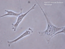
Using their new method, the researchers showed how the forces applied by fibroblast cells are actually distributed at the individual molecule level. “We found that each of the integrin receptors on the perimeter of cells is basically ‘feeling’ the mechanics of its environment,” Salaita says. “If the surface they feel is softer, they will unbind from it and if it’s more rigid, they will bind. They like to plant their stakes in firm ground.”

The integrin receptors on fibroblast cells, above, "are kind of beasts," Salaita says. "They apply relatively high forces in order to adhere to the extracellular matrix." NIH photo.

Each cell has thousands of these integrin receptors that span the cellular membrane. Cell biologists have long been focused on the chemical aspects of how integrin receptors sense the environment and interact with it, while the understanding of the mechanical aspects lagged. Cellular mechanics is a relatively new but growing field, which also involves biophysicists, engineers, chemists and other specialists.

“Lots of good and bad things that happen in the body are mediated by these integrin receptors, everything from wound healing to metastatic cancer, so it’s important to get a more complete picture of how these mechanisms work,” Salaita says.

The Salaita lab previously developed a fluorescent-sensor technique to visualize and measure mechanical forces on the surface of a cell using flexible polymers that act like tiny springs. These springs are chemically modified at both ends. One end gets a fluorescence-based turn-on sensor that will bind to an integrin receptor on the cell surface. The other end is chemically anchored to a microscope slide and a molecule that quenches fluorescence. As force is applied to the polymer spring, it extends. The distance from the quencher increases and the fluorescent signal turns on and grows brighter. Measuring the amount of fluorescent light emitted determines the amount of force being exerted. (Watch a video of the flexible polymer technique.)

Yun Zhang, a co-author of the Nature Communications paper and a graduate student in the Salaita lab, had the idea of using DNA molecular beacons instead of flexible polymers. “She was new to the lab and brought a fresh perspective,” Salaita says.

The molecular beacons are short pieces of lab-synthesized DNA, each consisting of about 20 base pairs, used in clinical diagnostics and research. The beacons are called DNA hairpins because of their shape.

The thermodynamics of DNA, its double-strand helix structure and the energy needed for it to fold are well understood, making the DNA hairpins more refined instruments for measuring force. Another key advantage is the fact that their ends are consistently the same distance apart, Salaita says, unlike the random coils of flexible polymers.

T cells are white blood cells whose receptors are focused not on adhesion, but on activities like identifying various peptides. Electron micrograph of a human T cell by NIAID/NIH.

In experiments, the DNA hairpins turned out to operate more like a toggle switch than a dimmer switch. “The polymer-based tension probes gradually unwind and become brighter as more force is applied,” Salaita says. “In contrast, DNA hairpins don’t budge until you apply a certain amount of force. And once that force is applied, they start unzipping and just keep unraveling.”

In addition, the researchers were able to calibrate the force constant of the DNA hairpins, making them highly tunable, digital instruments for calculating the amount of force applied by a molecule, down to the piconewton level.

“The force of gravity on an apple is about one newton, so we’re talking about a million-millionth of that,” Salaita says. “It’s sort of mind-bogging that that’s how little force you need to unfold a piece of DNA.”

The result is a tension probe that is three times more sensitive than the polymer probes.

In a separate paper, published in Nano Letters, the Salaita lab used the DNA-based probes to experiment with how the density of a substrate affects the force applied.

“Intuitively you might think that a less dense environment, offering fewer anchoring points, would result in more force per anchor,” Salaita said. “We found that it’s actually the opposite: You’re going to see less force per anchor.” The mechanism of sensing ligand spacing and adhering to a substrate appears to be force-mediated, he says. “The integrin receptors need to be closely spaced in order for the engine in the cell that generates force to engage with them and commit the force.”

Now the researchers are using the DNA-based tools they’ve developed to study the forces of more sensitive cellular pathways and receptors.

“Integrin receptors are kind of beasts, they apply relatively high forces in order to adhere to the extracellular matrix,” Salaita says. “There are lots of different cell receptors that apply much weaker forces.”

T cells, for example, are white blood cells whose receptors are focused not on adhesion but on activities like distinguishing a friendly self-peptide from a foreign bacterial peptide.

The Salaita lab is collaborating with medical researchers across Emory to understand the role of cellular mechanics in the immune system, blood clotting and neural patterning of axons. “Basically, our premise is that mechanics play a role in almost all biological processes, and with these DNA-based tension probes we’re going to uncover, measure and map those forces,” Salaita says.

Related:
Chemists reveal the force within you
Biochemical cell signals quantified for first time

Thursday, October 16, 2014

Ebola's backstory: How germs jump species

Fruit bats are associated with an array of deadly viruses, including Nipah, Ebola and Marburg. As the bats' habitat shrinks, the odds increase that bats will cross paths with humans, wild primates and other animals.

By Carol Clark
From Emory Medicine

While virologists study pathogens like Ebola by zooming in on them with an electron microscope, primate disease ecologist Thomas Gillespie climbs 100-foot trees in the tropical forests of Africa to get the big picture view. He tracks pathogens in the wild to learn how they adapt to changing environments and jump between species.

It is physically challenging work that often takes him into remote forests where the wildlife has not yet learned to fear people. A chimpanzee turned Gillespie into a human yo-yo while he was ascending a tree with a rope and harness. “Chimpanzees have 10 times the strength of a man and they can be curious and playful,” he says. “I once had an adult male chimpanzee grab my rope and bounce me up and down.”

Wild primates pose a special risk for zoonotic diseases—those transmissible from animals to humans—due to our close genetic relationship. HIV/AIDS and Ebola are the two most dramatic examples of diseases linked to wild primates, but many other viral, bacterial, fungal, and parasitic pathogens found in apes and monkeys are readily passed to humans.

“The bottom line is that the majority of emerging infectious diseases are coming from wildlife and most of that wildlife is in tropical forests,” says Gillespie, a professor in Emory’s Department of Environmental Sciences and the Rollins School of Public Health. “We can’t afford to just focus on one pathogen or one animal. It’s really important to get a general understanding of the interactions of different species, and how changes in the environment are driving zoonotic disease transmission.”


Gillespie is investigating undisturbed forests, as well as sites where logging and other human activity is under way. He gathers fecal and blood samples from people and animals for analysis while also scouring the forest floor and treetops to learn about the diversity of pathogens in the environment. The data can then be mapped spatially and over time to connect the dots of disease ecology.

For one ongoing study in Uganda, Gillespie and his collaborators are following primates in and around fig trees. The researchers hang out near these ancient forest giants, observing the tableau of life feeding amid the branches and on the ground below.

Fig trees are a keystone species of rainforest ecosystems. Climate change is playing havoc with the seasonal fruiting of other types of trees. But fig trees have co-evolved with specific pollinators—fig wasps—and due to their complex interaction, there is always a fig tree fruiting somewhere in the forest, providing a critical, consistent food source for fruit bats, primates, and ground dwellers like the bush duiker, a shy, dainty antelope that darts amid the forest shrubbery.

Fruit bats, associated with an array of deadly viruses including Nipah, Ebola, and Marburg, are especially specific in their diet. “They’re looking for ripe fruit,” Gillespie says, “and that’s a rare resource in the environment.”

And it’s becoming even rarer. Logging companies are cutting down huge swaths of African forests. Mining operations are moving into new terrain. Villages are expanding, and homes and food crops are eating into the wilderness. All these factors bump up the odds that fruit bats will be living near people, and that the bats will be joined by a variety of other animals while they are feeding from a tree.

“Most viruses can only last outside of a host for minutes or hours, not days,” Gillespie says, “but now we have this changing landscape of food availability. That raises the probability that a gorilla or chimpanzee will eat a piece of fruit that a bat has just defecated on, or has bitten into and discarded.”

Diseases and parasites could be transmitted in this manner. Ebola is one of the rare ones, extremely difficult to find, much less study, in the wild. But Ebola looms large in the public imagination because it is hemorrhagic, capable of causing massive bleeding, and because of its high fatality rate. It is also frightening because it is so mysterious, popping up out of the forest to kill voraciously then disappearing again for years.

The virus was first identified in 1976, following an outbreak in a remote hamlet of Zaire (now the Republic of Congo) near the Ebola River. Subsequent outbreaks have also been associated with forested backwaters and have quickly burned themselves out. That is, until the current outbreak in West Africa. Ebola has now made the leap from rural, forested regions to Africa’s urban areas, where many people live in crowded conditions with poor sanitation.

One of the biggest mysteries is where the virus has hidden between these outbreaks. Evidence of Ebola antibodies, and remnants of Ebola RNA, have been found in the blood of three species of fruit bats, making them prime suspects as an Ebola reservoir: An organism that can carry the pathogen without dying or even becoming sickened by it.

“Fruit bats are the best guess as to the reservoir, but until a live virus is found in their blood, we cannot be sure,” Gillespie says. “What we do know is that bats are an important part of the equation. And gorillas, chimpanzees, and some other animals, like the bush duiker, can get infected with Ebola.”

During the past decade, human Ebola outbreaks in Gabon and Congo have been accompanied by reports of gorilla and chimpanzee carcasses in surrounding forests, and epidemiological studies have connected encounters with dead gorillas, chimpanzees, and bush duikers to human cases.

“A hunter might find a dead gorilla in the forest,” Gillespie says, “and instead of saying, ‘I shouldn’t butcher this animal and eat it, it may have died of an infectious disease,’ he throws up his hands and says, ‘Thank you, God, for this gift!’ ”

Fruit bats are also hunted for food in many parts of Africa.

But you don’t have to be a hunter going deep into a forest to catch Ebola. Now that fruit bats are feeling the squeeze of fewer food sources, they may choose to roost under the eaves of a home, feasting on trees in the village orchard as children play below.

Widespread education about what is safe to eat and what is not, and how to identify animals that may have died from an illness, is becoming a vital part of preventing the spread of these diseases.

Just as people are encroaching on wilderness, pathogens are expanding their range into human habitats.

“We’re changing the environment in ways that may be promoting Ebola,” Gillespie says. “As the human population grows and the demand for resources pushes us into new areas, we’re going to see more diseases emerge. Anytime we alter a pristine natural system there are going to be unintended consequences.”

Photos: Thinkstock

Related:
Gorilla vet tracks microbes for global health
Mountain gorillas: People in their midst
Sanctuary chimps show high rates of drug-resistant staph

Thursday, October 9, 2014

Chemists uncover new role of a key base in organic synthesis

The collaboration of chemists from across three continents is a result of the Center for Selective C-H Functionalization (CCHF), an NSF National Center for Chemical Innovation headquartered at Emory. 

By Carol Clark

An international team of chemists has discovered a new piece to the puzzle of how a powerful base used in organic synthesis, cesium carbonate, plays a pivotal role during a catalytic reaction.

The research, published by the Journal of the American Chemical Society, was led by Jamal Musaev, a theoretical chemist at Emory University, and Ken Itami, an experimental chemist from Nagoya University in Japan. Sun Yat-Sen University in Guangzhou, China, also contributed to the findings.

Many organic chemistry reactions are acid/base reactions, involving the exchange of positively charged hydrogen atoms. Acids donate the positively charged hydrogen and bases accept it.

The current research focused on the use of cesium carbonate as a base. Cesium carbonate has recently been observed to accelerate a particular class of catalytic reactions, a phenomenon termed the “cesium effect.”

The use of cesium carbonate base and carboxylic acids co-catalysts have been shown to be critical in a number of recent carbon-hydrogen (C-H) bond functionalization reactions. The full story behind the impact of this base was previously not clear. It was known that the cesium base removed hydrogen protons, or scavenged acidic acid, from the solution, and was also involved in the exchange of ligands during a reaction, but these two factors did not explain the acceleration seen.

This recent work offers a new explanation. The researchers found that cesium base can generate an aggregate state: The molecules come together creating a cluster that is actually the starting point for the catalytic reaction, and not the discreet carboxylic acids and carbonate complexes as was previously thought.

“One-by-one, we are identifying key components of catalytic reactions and then putting them all together,” Musaev says. “It’s difficult work, but important, because the more your understand the reaction the better you can predict ways to modify it and control it.”

The findings about how the base acts in these reactions has the potential to impact the development of not just new C-H functionalization reactions, but the way that catalytic reactions in general are considered.

The collaboration of chemists from across three continents is a result of the Center for Selective C-H Functionalization (CCHF), an NSF National Center for Chemical Innovation headquartered at Emory. C-H functionalization holds the potential to make organic synthesis faster, simpler and greener, and could open up new ways to develop drugs and other fine-chemical products.

The CCHF encompasses 15 top research universities from across the United States, and recently expanded to include institutes in Asia and Europe. The global network forged by the CCHF brings together leading players from around the world, representing the range of specialties that will be required to make the critical breakthroughs needed to bring C-H functionalization into the mainstream of chemical synthesis.

Related:
Organic chemists now forming global bonds
NSF chemistry center opens new era in organic synthesis

Tuesday, October 7, 2014

Top 10 reasons to learn to make Stone Age tools

The Late Acheulean hand axe, going back about 500,000 years, "is the oldest technology that pretty much everyone agrees is unique to humans," says experimental archeologist Dietrich Stout.

By Carol Clark

Are you between the ages of 18 to 50, right-handed, and available for six hours per week? Emory experimental archeologists are looking for at least 20 healthy individuals willing to devote 100 hours over about four months to learn the art of making a Stone Age hand axe.

“We need novices who will really commit to learning this prehistoric craft,” says Dietrich Stout, an assistant professor of anthropology and head of the Paleolithic Technology Laboratory.

Nada Khreisheh will train participants.
Nada Khreisheh, a post-doctoral researcher in the lab, will train the participants to break and shape flint, a skill known as knapping, as part of a major, three-year archeology experiment to investigate the role of stone tools in human brain evolution, especially key areas of the brain related to language. For more details on how to apply, send her an email: nada.n.khreisheh@emory.edu.

In addition to attending tool-making training sessions, participants will undergo three magnetic resonance imaging (MRI) scans and eye-tracking experiments. And they will need to provide brief written feedback about their experiences following training sessions.

The ideal candidates to join the experiment would likely be curious about who we are as humans and where we came from. “If you’re not interested in trying to answer those questions, it might be hard to justify all the time and effort that will be involved,” Stout says.

If you meet the qualifying characteristics, here are 10 reasons you may want to apply to learn to make stone tools:

1. You will be making history.
“This is the first controlled, neuro-scientific study of real-world craft skill acquisition over time,” Stout says. “Our hypothesis is that the brain systems involved in putting together a sequence of words to make a meaningful sentence in spoken language overlap with systems involved in putting together a series of physical actions to reach a meaningful goal.”

Earlier studies, by Stout and others, have compared the brains of experienced knappers with novices. The results have all suggested that the part of the brain engaged in making a hand axe overlaps with areas associated with language. Longitudinal data, following people as they learn to master the art of making a hand axe, should provide a more definitive result, one way or the other. “This is a much more focused and rigorous test than any previous study,” Stout says.

At the same time, the researchers hope to develop the first systematic model for describing the syntax of natural human action. “We’re proposing a method to break actions down into ‘phrases’, quantify their ‘grammatical’ structure and relate this directly to processing in the brain,” Stout says. “Although we’re using the domain of tool knapping, the same method may apply more broadly to any complex series of actions.”

2. You will be making prehistory. 
The hand axe represents a pivotal point in prehistory and an ideal technology to hone in on key ways that we shifted from more ape-like hominids into full-fledged humans, Stout says. Simple Oldowan stone flakes are the earliest known tools, dating back 2.6 million years, before the human family emerged. The Late Acheulean hand axe, going back 500,000 years, embodies a much higher level of refinement and standardization.

 “You see a clear increase in complexity in the hand axe,” Stout says. “It’s the oldest technology that pretty much everyone agrees is unique to humans.”

The stone tool experiment has its own logo, designed by Khreisheh.

3. You can get in touch with your Acheulean roots.
Stout says doing knapping himself gives him a unique connection to the past. “I can pick up a stone tool from an archeological site and see things that are so familiar to my own experience: A flake taken off here and there, and then there is a ding where the person who was knapping the stone tried something that didn’t work,” he says. “I’ll get this sense of what that prehistoric person might have been feeling. It gives you goose bumps.”

4. It is an interesting challenge. 
“Many people expect it to be easy, because it’s ancient technology,” Stout says, but it’s actually challenging to chip out the lens-shaped cross-section of the hand axe, and thin down its edges to expert sharpness. “A lot of self-control is involved in flint knapping. You have to not get frustrated and just start banging on the rock,” he says.

5. It’s fun, once you get the hang of it.
“I really enjoy it, it’s kind of additive,” says Khreisheh, who began knapping a decade ago as part of her research at the University of Exeter, England. Greensand silicate, found near where she grew up, is her favorite material to work with and she has amassed a large collection of her hand-made tools. “When I was packing up my stuff to move to Atlanta, I had so many rocks it was just ridiculous.”

6. It may give you an edge.
“It’s not just another skill, it will really set you apart,” Khreisheh says. “When I tell people that I’m a flint knapper, they usually have no idea what that means but they are always interested in hearing about it.”

Dietrich Stout surveys his stash of flint stones near the Anthropology building.

7. You will never have to knap alone. 
If you decide to turn it an ongoing hobby, you can tap into an established community of knappers. They have conferences, publications and even exhibitions of lithic art. “Some of the better pieces are like sculpture,” Stout says.

8. It will change the way you look at rocks.
“It enriches your vision,” Stout says. “Other people may just see a rock, but you see all kinds of features in a stone, like, ‘This is where it would break if I hit it.’ The other day when I was walking on campus past some gravel landscaping, I thought, ‘That looks like it would make a good hammer stone. I could really use that one.’”

9. Stone tools have practical uses. 
You could not ask for a more impressive paperweight than a hand axe that you made yourself.

“I grab one of these things when I want to open a box,” says Stout, waving at the array of stone flakes spread out in his lab.

“One of my advisors made a lemon squeezer out of blade cores,” Khreisheh adds.

Another dedicated archeologist actually elected to have major surgery done with obsidian blades, rather than steel scalpels, to demonstrate to his students that stone tools are more technologically advanced than many people realize.

10. Understanding neural systems may lead to broader applications.
“Any insights into how people understand physical activity and language may lead to new ways to help people with brain damage or language difficulties,” Stout says. He hopes the tool-making experiment will benefit a range of neuroscience research.

“Neuroscientists tend to focus on the organism itself,” he says, “but humans are immersed in material culture. Much of our identity and experience is dictated by our stuff. As experimental archeologists, we bring a deep understanding of technology, culture and tools to the study of the human brain.”

The project is funded by the National Science Foundation and the John Templeton Foundation, through a program designed to integrate science across disciplines.

All photos by Bryan Meltz, Emory Photo/Video.

Related:
Brain trumps hand in Stone Age tool study
Hominid skull hints at later brain evolution
A brainy time traveler

Wednesday, October 1, 2014

Former Pres. Carter celebrates 90th birthday with butterfly garden dedication

Garden party: On his 90th birthday, former U.S. President Jimmy Carter and his wife, Rosalynn, celebrated in their new pollinator garden. Among the guests was Emory evolutionary ecologist Jaap de Roode, bottom right, and his children Jakob and Ella.

By Megan McRainey

Former U.S. President Jimmy Carter celebrated his 90th birthday at The Carter Center today with a tour of a new butterfly garden created in his honor.

The Jimmy and Rosalynn Carter Pollinator Garden created with the help of Emory University evolutionary ecologist Jaap de Roode, is filled with flowers and plants native to Georgia and is part of the Rosalynn Carter Butterfly Trail, developed by the former first lady to draw attention to the plight of diminishing numbers of migrating monarch butterflies. The garden is certified by Monarch Watch as an official monarch way station, and is listed as a certified wildlife habitat of the National Wildlife Federation.

De Roode and undergraduate students from his lab worked with volunteers from Trees Atlanta to provide seedlings for the garden and to plant them over the summer. They will continue to help monitor and maintain the plants.

The garden features two species of milkweed, the host plants monarch caterpillars need to complete their life cycle, and a variety of plants that can host Georgia’s state butterfly, the Tiger Swallowtail. An array of nectar plants also will appeal to other pollinator species, particularly bees and wasps, and to birds. Visitors are welcome to drop by the garden, which also features two Japanese-style arbors made by local artist Jesse Reep.

De Roode’s lab is one of only a few labs in the world devoted to the study of monarch butterflies and their parasites. In 2010, the lab discovered that monarchs use the toxic chemicals in certain species of milkweed to rid themselves of harmful parasites.

Jaap de Roode and his children at work in the garden last June, along with students from his lab, including, from left: Michelle Tsai, Kevin Hoang, Camden Gowler and Itai Doron.

Nature published de Roode's latest paper on monarchs today, involving the genomic analysis of monarch butterflies from around the world. The researchers traced the ancestral lineage of monarchs to a migratory population that likely originated in the southern United States or Mexico, instead of South America, as was previously hypothesized.

Every year, millions of monarchs from Canada and the United States fly south to overwinter in Mexico. Due to declining amounts of milkweed, from pesticide use and development of land, the number of monarchs making the migration has dropped dramatically, from an estimated180-900 million in 1996-1997 to an estimated 6.7-33 million in 2013-2014.

“The whole migrating population could be gone over the next decade,” de Roode says. “It’s an amazing natural phenomenon in danger of disappearing.”

Initiatives such as the Rosalynn Carter Butterfly Trail can help reverse the trend, he adds, and encourage individual gardeners to lend more support to monarchs by planting milkweed. The trail, which begins in Plains, Georgia, now includes registered sites extending as far as Canada.

Born on Oct. 1, 1924, Jimmy Carter served as the 39th President of the United States from 1977 to 1981 and joined Emory’s faculty in 1982, the same year he established The Carter Center. He is the University Distinguished Professor at Emory.

Related:
Pumping wings: Muscles make migrating monarchs unique

Pumping wings: Muscles make migrating monarchs unique

Genes related to muscle function distinguish the monarchs that migrate over thousands of miles.

By Carol Clark

A major study that sequenced the genomes of monarch butterflies from around the world identified a gene related to muscle function that appears central to the monarch’s spectacular annual mass migration across North America.

The findings, published in Nature October 1, also traced the evolutionary origins of monarchs to North America, instead of South America as was previously hypothesized, and identified a gene related to the butterfly’s distinctive orange-and-black coloration.

“Our findings add several interesting twists to our understanding of these iconic insects,” says Jaap de Roode, an evolutionary ecologist at Emory and a co-author of the paper.

Evolutionary geneticist Marcus Kronforst from the University of Chicago led the sequencing of the genomes of 101 monarchs, which involved scientists from five other universities, in addition to Emory. De Roode, who runs one of a handful of labs in the world focused on monarch butterflies, assisted in the design of the project and the analysis of the results.

The researchers traced the ancestral lineage of monarchs to a migratory population that likely originated in the southern United States or Mexico. The evolutionary tree created from the sequencing showed that the monarch’s current worldwide distribution appears to stem from three separate dispersal events – to Central and South America; across the Atlantic; and across the Pacific. In all three cases, the butterfly independently lost its migratory behavior.

In addition to better muscle function, migrating monarchs have more efficient metabolisms than populations of monarchs that have not evolved the ability for long migrations. Photo by Jaap de Roode.

The monarch’s North American origin runs counter to a long-standing hypothesis that the butterfly originated from a non-migratory tropical species.

“Previously, it was widely thought that after spreading from the tropics through North America, the evolution of migration enabled monarchs to fly south and survive the winter,” de Roode says. “It turns out that we had that upside down.”

To better understand the genetic basis for the butterfly’s migratory behavior, the researchers compared the genomes of migratory and non-migratory monarch populations from around the world. A disparity between the two groups among genes related to muscle function stood out, including one in particular: collagen IV alpha-1. The migratory butterflies expressed greatly reduced levels of this gene, which is involved in muscle function.

Humans have a similar gene that is associated with a muscle disease known as myopathy, de Roode notes.

“The data clearly show that muscles are the main thing that enables monarchs to migrate over thousands of miles,” he says. “We also found that the migrating monarchs have much more efficient metabolisms.”

By comparing the genome of mutant white monarchs, found in Hawaii, with other monarchs the researchers identified a gene clearly correlated with the butterfly’s beautiful orange color.

A similar gene found in mice, myosin 5a, is associated with a dilute phenotype: Instead of the black fur, mice with a mutation in this gene are light brown or beige. “Myosin 5a is a gene that regulates transport of pigment within a cell, moving color to a hair shaft in the case of mice, or apparently to scales in the case of monarchs,” de Roode says. The gene has never before been implicated in insect coloration, he adds.

In 2010, de Roode’s lab discovered that monarchs use a toxic chemical in certain species of milkweed to rid themselves of harmful parasites, providing some of the best evidence that animals use medication.

“Monarchs are a fascinating system to study,” he says.

“There is so much that we don’t yet know about these insects, and we may never know because they are now in decline.”

Monarch larvae feed on milkweed, which is becoming rarer due to pesticide use and development of land. As a result, the number of monarchs making the annual migration from Canada and the United States to Mexico has dropped dramatically: From an estimated 180 million-900 million in 1996-1997 to an estimated 6.7 million-33 million this past year.

“The whole migrating population could be gone over the next decade,” de Roode says. “It’s an amazing natural phenomenon in danger of disappearing.”

De Roode and his students helped create the Jimmy and Rosalynn Carter Pollinator Garden at The Carter Center, which opened October 1 in honor of President Carter’s 90th birthday. The garden is filled with milkweed and flowers and plants native to Georgia and is part of the Rosalynn Carter Butterfly Trail, developed by the former first lady to draw attention to the plight of the monarchs.

Credits: Top and bottom photos via ThinkStock; center photo by Jaap de Roode.

Related:
Monarch butterflies use drugs
Mystery of monarch migration takes new turn