Showing posts with label Chemistry. Show all posts

Monday, March 16, 2026

Turning Over a New Leaf in Analyses of Natural Products

Emory graduate student William Crandall loves working at the nexus of nature and cutting-edge technology. (Photo by Tharanga Samarakoon)

Scientists developed a new way to help understand what happens in the body when people consume a plant product and the many chemicals it contains. The American Chemical Society’s Journal of Natural Products published the method to quickly analyze the effects of a natural product, developed at Emory University. 

As a test case, the paper focused on biotransformation of chemicals from the kratom plant by human liver cells in a laboratory dish. The researchers developed an automated method — based on high-resolution mass spectrometry and molecular network mapping — to gain a detailed, big-picture view of the resulting metabolites, or chemicals produced. 

The new, streamlined methodology can be broadly applied to nutrition and dietary supplement research, filling a critical gap in the field. 

“Plants evolved extraordinarily complex chemical defenses and signaling systems,” says Cassandra Quave, co-senior author of the study and professor of dermatology at Emory School of Medicine and the Center for the Study of Human Health. “Our new approach in molecular mapping gives us a way to follow how that chemical complexity is reshaped by human metabolism.” 

“Our technique does not just look at how one compound in this plant is metabolized,” adds William Crandall, first author of the study and a PhD student of molecular and systems pharmacology in Emory’s Laney Graduate School. “It shows how dozens of compounds are metabolized at one time.”

“This method marks a major, transformative step in natural products research,” says Dean Jones, co-senior author of the paper and professor in Emory School of Medicine. “A process that used to require years of work now takes just days.”


Related:


Wednesday, January 7, 2026

Unlocking design secrets of deep-sea microbes

"The molecular study of proteins is rapidly expanding as the technology supporting the field keeps advancing," says Vincent Conticello. "You're only limited by your interest and your imagination." (Photo by Carol Clark)

The microbe Pyrodictium abyssi is an archaeaon — a member of what’s known as the third domain of life — and an extremophile. It lives in deep-sea thermal vents, at temperatures above the boiling point of water, without light or oxygen, withstanding the enormous pressure at ocean depths of thousands of meters. 

A biomatrix of tiny tubes of protein, known as cannulae, link cells of Pyrodictium abyssi together into a highly stable microbial community. No one knew how these single-celled microbes accomplished this feat of extreme engineering — until now. 

A study using advanced microscopy techniques reveals new details about the elegant design of the cannulae and the remarkable simplicity of their method of construction. Nature Communications published the work, led by scientists at Emory University; the University of Virginia, Charlottesville; and Vrije Universiteit Brussel in Belgium. 

The discovery holds the potential to inspire innovations in biotechnology, from the development of new “smart” materials to nanoscale drug delivery systems. 

“Not only are the cannulae strong enough to endure extreme conditions, they’re beautiful,” says Vincent Conticello, Emory professor of chemistry and co-senior author of the paper. “To me, they resemble columns from the classical architecture of ancient Greece or Rome,” he adds, citing their fluted edges and precise regularity.

Read the full story here.

Related:

Emory chemists invent shape-shifting nanomaterial

Tuesday, September 23, 2025

New methods expand access to molecules key to human health

"I love solving problems, the more challenging the better," says San Pham, first author of the paper. Senior author is Frank McDonald, Emory professor of chemistry and Pham's PhD advisor.

A new approach to an established reaction boosts the ability to synthesize vinylic ethers — key building blocks for many molecules that are important to human health. The American Chemical Society’s Organic Letters published the breakthrough, made by chemists at Emory University. 

“Our method is easy to reproduce and is based on widely available and inexpensive compounds,” says San Pham, an Emory PhD candidate and first author of the paper. “We can apply this method to make multiple natural products, including novel vinylic ethers.” 

Her research improves the reliability, yield and generality of what is known as the Chan-Evans-Lam reaction. These enhancements greatly expand the reaction’s potential for the synthesis of complex, biologically active compounds for drug research.


Related:


Tuesday, April 15, 2025

New AI tool set to speed quest for advanced superconductors

Xu Chen, an Emory PhD student of theoretical chemistry, is first author of the paper. He says the team was inspired by the image-recognition training used for self-driving cars to create a powerful machine-learning framework.

Using artificial intelligence shortens the time to identify complex quantum phases in materials from months to minutes, finds a new study published in Newton. The breakthrough could significantly speed up research into quantum materials, particularly low-dimensional superconductors. 

The study was led by theorists at Emory University and experimentalists at Yale University. Senior authors include Fang Liu and Yao Wang, assistant professors in Emory’s Department of Chemistry, and Yu He, assistant professor in Yale’s Department of Applied Physics. 

The team applied machine-learning techniques to detect clear spectral signals that indicate phase transitions in quantum materials — systems where electrons are strongly entangled. These materials are notoriously difficult to model with traditional physics because of their unpredictable fluctuations. 

“Our method gives a fast and accurate snapshot of a very complex phase transition, at virtually no cost,” says Xu Chen, the study’s first author and an Emory PhD student in chemistry. “We hope this can dramatically speed up discoveries in the field of superconductivity.” 

One of the challenges in applying machine learning to quantum materials is the lack of sufficient high-quality experimental data needed to train models. To overcome this, the researchers used high-throughput simulations to generate large amounts of data. They then combined these simulation results with just a small amount of experimental data to create a powerful and efficient machine-learning framework.

Read more about the discovery.

Related:

Chatbot opens computational chemistry to nonexperts

Monday, April 7, 2025

Chatbot opens computational chemistry to nonexperts

The researchers hope their pioneering work to democratize computational chemistry will inspire similar initiatives across the natural sciences. (Liu Group)

Advanced computational software is streamlining quantum chemistry research by automating many of the processes of running molecular simulations. The complicated design of these software packages, however, often limits their use to theoretical chemists trained in specialized computing techniques. 

A new web platform developed at Emory University overcomes this limitation with a user-friendly chatbot. The chatbot guides nonexperts through a multistep process for setting up molecular simulations and visualizing molecules in solution. It enables any chemist — including undergraduate chemistry majors — to configure and execute complex quantum mechanical simulations through chatting. 

The free, publicly available platform — known as AutoSolvateWeb — operates primarily on cloud infrastructure, further expanding access to sophisticated computational research tools. 

The journal Chemical Science published a proof-of-concept for AutoSolvateWeb, which marks a significant step forward in the integration of AI into education and scientific research.

Related:


Wednesday, March 5, 2025

Atlanta Science Festival set to entertain, inspire and engage all ages

The festival culminates Saturday, March 22, in "Exploration Expo," a day-long celebration in Piedmont Park. Demonstrations by Emory chemist Douglas Mulford are among the perennial favorites.

By Carol Clark

Atlanta Science Festival returns March 8-22, with more than 100 events throughout the metro area, inviting the public to join fun, interactive and educational experiences. The acclaimed city-wide celebration, one of the largest of its kind in the country, showcases the myriad science, technology engineering and mathematics (STEM) innovations happening in Atlanta, including at Emory. 

“Not only does the Atlanta Science Festival spotlight the wonder of science in its various forms, we strive to do so by curating a two-week experience that’s as exciting and intriguing as possible,” says Meisa Salaita, executive co-director of Science ATL, the non-profit organization that engineers the festival. “We want to open minds, educate, inspire, entertain, and spark the interest of the scientists of tomorrow.” 

Now in its 12th year, the Atlanta Science Festival was co-founded by Emory, Georgia Tech and the Metro Atlanta Chamber. 

Members of the Emory community will help participants experience the wonders of science through spectacles like the chemistry of fireballs, a musical entertainment combined with a biology talk on the surprising abilities of animals to use medicine, a walking tour of campus science landmarks, a behind-the-scenes look at the latest advances in healthcare technology and much more. 

Creative events to engage participants with technology include “Data Poetics,” which will combine slam poetry and computer science on Thursday, March 13 at 7 p.m. at the Supermarket event space in Atlanta. The introductory workshop in how to use software to visualize data and add power to poetic expression will be co-hosted by Emily Wall, Emory assistant professor of computer science, Keke Wu, Emory postdoctoral researcher, and W. J. Lofton, an Atlanta poet. 

The idea for the event grew out of an Emory class that Wall and Lofton co-taught as part of the Emory Arts and Social Justice Fellows program, which pairs faculty with local artists to explore how creative thinking and artistic expression can inspire change. Their class was so successful that the duo wanted to introduce the concept to the wider public. 

Participants will write a data-driven poem about a social issue affecting Atlanta and then amplify their message through information visualizations. “Many people think of computer science as intimidating and too ‘mathy’ to be interesting,” Wall says. 

That attitude often changes when people learn simple ways to directly apply computer science to better communicate a human problem, she adds. “We want to give artists another tool, a way to make their art even more compelling.”

Tuesday, February 11, 2025

Plant extract inspires new chemistry and new early lead against triple-negative breast cancer

The extract that inspired the research comes from Curcuma phaeocaulis, a flowering plant in the ginger family. (Wagner Campelo / Alamy Stock Photo)

Chemists at Emory University invented a reaction to streamline the total synthesis of a compound, phaeocaulisin A, extracted from a plant used for centuries in traditional Chinese medicine. 

In laboratory dish experiments conducted with biologists at Winship Cancer Institute of Emory University, the researchers showed the compound’s efficacy against HER2-positive breast cancer cells and triple-negative breast cancer cells. An analogue of the compound the chemists constructed boosted this efficacy. 

“We not only efficiently replicated a complex natural product,” says Mingji Dai, Emory professor of chemistry. “We also improved upon it by turning it into a more potent compound.” 

The Journal of the American Chemical Society published the work, led by Dai and Yong Wan, professor of pharmacology and chemical biology at Emory School of Medicine and director of basic research for the Glenn Family Breast Center at Winship Cancer Institute.


Related:

Wednesday, November 13, 2024

Chemists showcase power of pathbreaking method to make complex molecules

"We've had tremendous impact on developing C-H functionalization as both an academic discipline and for industry applications," says Emory chemist Huw Davies, who brought researchers from 15 universities together under the umbrella of the NSF Center for C-H Functionalization.

Chemists synthesized a highly complex natural molecule through a revolutionary strategy of functionalizing normally inert carbon-hydrogen (C-H). Science published the breakthrough led by chemists at Emory University and Caltech. 

The work is the most dramatic example yet of a sequence of C-H functionalization reactions selectively transforming low-cost materials into complex building blocks of organic chemistry. Ten of the steps involved in their synthesis of cylindrocyclophane A — a natural compound with antimicrobial properties — involved C-H reactions. 

“It’s by far the most complex natural product we have made using our method,” says Huw Davies, Emory professor of chemistry and co-corresponding author of the paper. “This is a game changer. We’re doing chemistry on C-H bonds that formerly would have been considered as unreactive. And we’ve shown how we can orchestrate a suite of 10 C-H functionalization steps, targeting a single C-H bond at a time in a specific sequence.” 

“This work moves the field forward by showing the power of C-H functionalization,” adds Brian Stoltz, professor of chemistry at Caltech and co-corresponding author of the paper. “It will open people’s eyes to the possibilities of using these very selective and unusual transformations in a really complex setting.” 

First author is Aaron Bosse, who did the work as an Emory PhD student. Bosse has since graduated and is now a medicinal chemist at Takeda Pharmaceuticals in Cambridge, Massachusetts.


Tuesday, October 29, 2024

Bacterial pathogen shows alarming resistance to common cleaners, chemists discover


A new study reveals widespread resistance of a major bacterial pathogen to the active ingredients in cleaning agents commonly used in hospitals and homes. 

The American Chemical Society Infectious Diseases published the research led by chemists at Emory University. It demonstrates the surprising level of resistance to cleaning agents of multidrug-resistant Pseudomonas aeruginosa, a pathogen of particular concern in hospital settings. 

The study also identifies biocides that are highly effective against P. aeruginosa, including a novel compound developed at Emory in collaboration with Villanova University. The researchers describe how these biocides work differently than most disinfectants currently in use. 

“We hope our findings can help guide hospitals to reconsider protocols for the sanitation of patient rooms and other facilities,” says William Wuest, Emory professor of chemistry and a senior author of the study. “We also hope that our findings of a new mechanism of action against these bacterial strains may help in the design of future disinfectant products.” 

First authors of the study are Christian Sanchez (who did the work as an Emory PhD student in chemistry and, following graduation, joined the faculty at Samford University) and German Vargas-Cuebas, an Emory PhD candidate in microbiology through Laney Graduate School. 

“Resistance of pathogens to cleaning agents is an area that’s often overlooked,” Vargas-Cuebas says, “but it’s an important area of study, especially with the rise in antibiotic-resistant pathogens worldwide.”


Related:


Friday, March 29, 2024

A new estimate of U.S. soil organic carbon to improve Earth system models

"To understand how soil carbon will change under a changing climate, we first need accurate estimates of current soil organic carbon levels and the key factors that influence them," says Emory environmental scientist Debjani Sihi, senior author of the study.

By Carol Clark

Soil contains about twice as much carbon as the atmosphere and plants combined. It is a major carbon sink, capable of absorbing more carbon dioxide from the atmosphere than it releases. Management of soil carbon is key in efforts to mitigate climate change, in addition to being vital to soil health and agricultural productivity. 

Measuring soil carbon, however, is a painstaking, expensive process. Samples must be dug from the ground and sent to a lab for analysis, making upscaling measurements on a large spatial scale challenging. 

Now environmental scientists have combined field-level data with machine-learning techniques to estimate soil organic carbon at the U.S. continental scale. The Journal of Geophysical Research — Biogeosciences published the new soil organic carbon estimate, which improves the overall estimate for the United States and gives new insights into the effects of environmental variables on soil organic carbon. 

“There is growing recognition that soil organic carbon is important and that we should invest in building it up through sustainable land management practices,” says Debjani Sihi, senior author of the study and assistant professor of environmental sciences at Emory University. “Our estimate is more accurate than existing estimates and provides a better benchmark to guide policymakers and land managers in adopting climate-smart practices.” 

Land is far more efficient than the ocean at retaining carbon, Sihi notes, and offers one possible nature-based solution to help mitigate climate change. “We could potentially create conditions,” she explains, “that are favorable for soil to capture carbon dioxide from the atmosphere and lock it there for a really long time — over millennia.” 

Sihi is a biogeochemist who studies environmental and sustainability issues at the nexus of soil and climate. 

First author of the current paper is Zhuonan Wang, a former postdoctoral fellow in Sihi’s lab who is now at Colorado State University. 

Digging into soil data 

Soil organic carbon is made up of plant and animal matter in various states of decomposition. While inorganic carbon is also found in the soil in the form of carbonate minerals, organic carbon is usually the largest proportion and the most important driver of soil biology and quality. 

The U.S. Department of Agriculture maintains the National Cooperative Soil Survey soil characterization database. This data was gathered over decades both by walking over the land and observing and by digging up core samples and sending them to laboratories for analysis. Measuring soil organic carbon, for example, requires digging a core to the root zone, about 30 centimeters deep to obtain a topsoil profile and until the core hits bedrock to obtain an entire soil profile. 

Soil sampling is done in other parts of the world as well. The International Soil Organic Carbon Network encompasses more than 430,000 soil profiles, drawn from across the globe. Scientists use such data to create “soil maps,” or estimates of soil characteristics in various regions. One well-known soil map is the Harmonized World Soil Database, developed by the United Nations’ Food and Agricultural Organization and collaborators. Another is SoilGrids, supported by the International Soil Reference and Information Center in the Netherlands. 

Significant inconsistencies exist in the estimates of soil organic carbon within both the Harmonized World Soil Database and SoilGrids. Sihi and her team set out to see if they could resolve these inconsistencies within the United States estimates by finding more effective ways to scale up the soil-sampling data. 

The researchers divided the United States — including all 50 states and Puerto Rico — into 20 different regions and created machine-learning models for each region. They obtained nearly 50,000 soil samples, ranging from 30 centimeters to one meter deep, from across these regions. They built their algorithms using these data samples for soil organic carbon, matched to precise geographic information system locations. 

They also drew from additional open-source data to feed their models with 36 environmental variables, including details about the climate, topographical features of the land, biogeochemical properties of the soil and the amount of vegetation on the landscape. 

A better benchmark for modeling Earth systems 

The results showed that the new method provided more accurate estimates than both the Harmonized World Soil Database and SoilGrids for the top 30 centimeters of soil, where the most biologically active soil organic carbon tends to be concentrated. 

The new method also revealed how the effects of environmental variables on soil organic carbon vary across regions. While climate was the most common predictor of soil organic carbon across most of the regions, the vegetation index tended to be more important in the arid areas of the southwest. Elevation was a key variable in regions that were mountainous or included a major river delta. 

The researchers hope that others will apply their approach to other countries and continents where enough on-the-ground data is available. 

“The beauty of our approach is that it gives us the power to identify regions with high uncertainty in our estimates and that helps us to guide future sampling efforts,” Sihi says. 

Considering environmental variables also increases the new model’s flexibility as global temperatures rise due to climate change, causing soils to warm and alter rainfall patterns. It remains unclear, Sihi notes, if soils will continue to serve as a carbon sink or transform into a carbon source. 

“To understand how soil carbon will change under a changing climate, we first need accurate estimates of current soil organic carbon levels and the key factors that influence them,” Sihi says. “Our new estimate is a step toward getting more accurate baseline data to improve Earth system models for climate change.” 

Co-authors of the new estimate include Jitendra Kumar (Oak Ridge National Laboratory), Samantha Weintraub-Leff (National Ecological Observatory Network), Katherine Todd-Brown (University of Florida) and Umakant Mishra (Sandia National Laboratories). 

The work was supported by the National Science Foundation and the U.S. Department of Energy.

Related:


Wednesday, March 6, 2024

Atlanta Science Festival returns to inspire discovery for all ages

A middle-school student experiences an Emory chemistry lab during a recent community outreach event. 

The Atlanta Science Festival returns March 9 to 23, inviting curious kids and adults to explore all things science, technology, engineering and mathematics (STEM). Experts in these fields — including many members of the Emory community — will serve as educational guides for more than 150 interactive events. 

“The Atlanta Science Festival aims to bring the community together through their shared love of science,” says Meisa Salaita, co-founder and co-executive director of Science ATL, the engineers of the festival. “Through these events, we hope to inspire and empower the next generation to pursue their dreams.” 

Participants can take a crash course on the basics of AI, create an herbarium of medicinal plants, go into the field with researchers studying microplastic pollution in a stream, take a behind-the-scenes tour of the latest advances in healthcare technology and even get a taste of the physics of cheese making. 

Now in its 11th year, the Atlanta Science Festival was co-founded by Emory, Georgia Tech and the Metro Atlanta Chamber. 

“We have grown into a mainstay of Atlanta,” says Salaita, noting that many of the events fill up quickly. “The festival is something that people look forward to every spring.” 

Tuesday, December 12, 2023

New tool to analyze blood platelets holds major medical potential

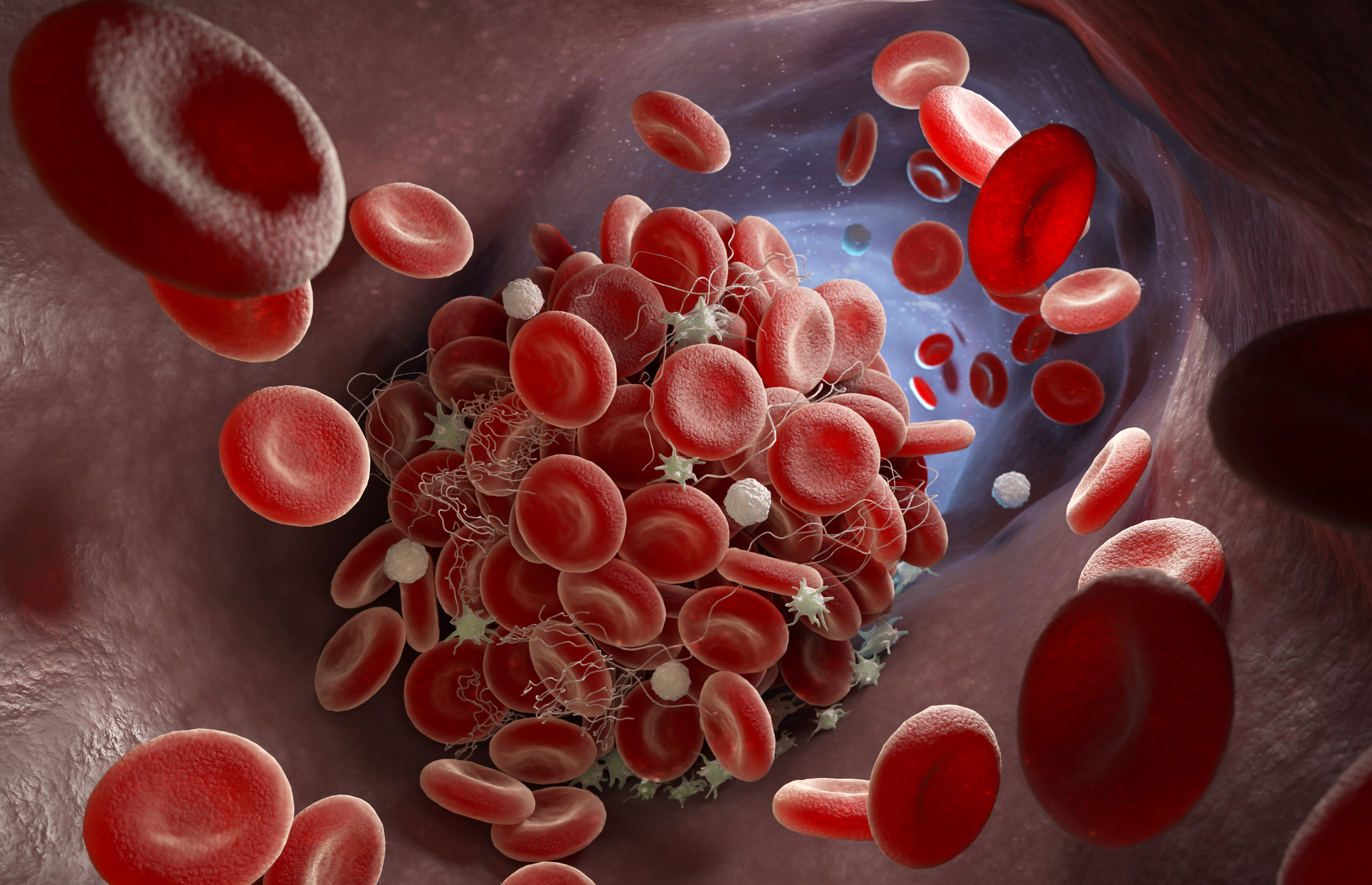
Graphic image shows a blood clot forming in an artery. The white, spikey platelets are amid red and white blood cells. Activated platelets use their spkes like "arms" to grip onto one another and to stringy chains of proteins in the blood called fibrin, forming clumps that also bind up red and white blood cells.

By Carol Clark

A novel technique to test platelet function within a person’s blood sample is faster, easier and more precise than methods currently in use, an experimental study shows. 

Nature Biomedical Engineering published the research, led by scientists at Emory University. The researchers demonstrated the proof-of-concept for the technique, which provides the first detailed look at the molecular forces generated by activated platelets in patient blood samples. 

The study results show that the technology holds the potential to assess the effects of antiplatelet drugs on individuals and to gain a clearer picture of bleeding risks for patients undergoing cardiopulmonary bypass surgery. 

The technique requires only about a drop of blood to run tests, compared to the tablespoon needed for current assays. This ultrasensitivity may make the technology a valuable tool for the diagnosis of babies suffering from rare, congenital platelet disorders. 

The breakthrough is based on synthetic-DNA tension probes developed more than a decade ago in the laboratory of Khalid Salaita, professor in Emory’s Department of Chemistry and in the Wallace H. Coulter Department of Biomedical Engineering at Emory and Georgia Tech. 

‘A scientist’s dream’ 

The tension probes can detect cellular forces on the magnitude of just a few piconewtons, or about a billion times less than the weight of a paper clip. The researchers found a way to amplify the signal of the probes by tapping the power of an enzyme known as CRISPR-associated 12a. The mechanical signal is then detected using a plate-reader, a tool already routinely used in clinical testing. 

“This project started out of basic curiosity,” says Salaita, co-corresponding author. “We wanted to know whether we could measure the tiniest forces exerted by cells. It’s exciting that we are now building on this basic curiosity to develop diagnostic tools to help patients. It’s a scientist’s dream.” 

First author of the paper is Yuxin Duan, an American Heart Association postdoctoral fellow in the Salaita lab. 

Roman Sniecinski, a professor in Emory School of Medicine’s Department of Anesthesiology and a leading expert in the field of perioperative coagulation, is co-corresponding author of the paper.

“Platelet function in general is important and yet the current tools that we have to measure it are relatively primitive,” Sniecinski says. “This new technique offers an easier, faster and cheaper way to measure platelet function, while also providing us with key information that we didn’t have before.” 

Co-authors include: Fania Szlam, a senior associate in the Sniecinski lab; Yuesong Hu, a graduate student in the Salaita lab; Renhao Li, a professor in Emory School of Medicine’s Department of Pediatrics, Hematology/Oncology; Wenchun Chen, a postdoctoral fellow in Emory School of Medicine; and Yonggang Ke, an associate professor in the Coulter Department of Biomedical Engineering at Emory and Georgia Tech. 

The importance of platelets 

Platelets are colorless, disc-shaped blood-cell fragments whose job is to bind at the site of an injured blood vessel to stop the bleeding. In some cases, however, platelets may not function optimally. When platelets are weak, or less active than optimal, the blood may not clot properly leading to uncontrolled bleeding. But if platelets are “hyperactive” they may become too sticky and cause spontaneous blood clots that can lead to heart attack or stroke. 

Regulating platelet function is especially critical to people at higher risk for some conditions. Antiplatelet drugs, such as clopidogrel, ticagrelor and even aspirin, are among the most commonly prescribed medications in the United States. In some patients, however, these drugs may not work well and adjustments in doses or changing to another drug might better help prevent heart attacks. 

During cardiac surgery, platelet function becomes even more dysregulated. The operating-room team must perform a balancing act of making blood not clot during cardiopulmonary bypass, then using procoagulant interventions, including transfusions of platelets, to stop the bleeding when the surgical procedure is finished. This can be difficult because the use of the cardiopulmonary bypass machine can stress and weaken blood platelets. 

“For decades, people have written in the scientific literature about this problem of platelet dysfunction during cardiac surgery,” Sniecinski says, “but it’s really difficult to measure it with the tools that we’ve been using. And since we haven’t been able to measure platelet function well, that’s made it difficult to study it in effective ways.” 

‘A small part of the picture’ 

Aggregometry is a standard tool currently used to assess platelet function. It measures the speed and degree at which platelets in a blood sample clump together, or aggregate. 

“This data provides only a small part of the picture of platelet function and it’s not the most interesting part,” Sniecinski says. 

When a platelet gets activated, he explains, it changes its morphology and grows tiny pseudo “arms.” Platelets use these arms to grip onto chains of proteins in the blood called fibrinogen to form clots. 

“Aggregometry tells you that platelets are clumping together,” Sniecinski says. “But it doesn’t tell you about their level of activation — the amount of force they’re using to hold on to other coagulation proteins, as well as each other.” 

Amplifying the signal 

The Salaita lab is a leader in visualizing and measuring the mechanical forces applied by cells using tension probes made from synthetic strands of double-stranded DNA tethered to a surface. 

The double-strands of DNA can be programmed to bind to platelet cells. When the cells bind and apply force to the anchored DNA, the DNA splits into two strands, leaving one strand stuck to the surface. The resulting physical tug is converted into a fluorescent signal. 

A major challenge to reading this signal, however, is that these physical tugs are faint, fleeting and infrequent. They require a microscope to detect them. 

During the COVID-19 pandemic , the enzyme CRISPR 12a, or Cas12a, came to the fore as a diagnostic tool for SARS-CoV-2 virus. Bacteria use Cas12a to defend against phages, or viruses that attack bacteria. The Cas12a enzyme can be loaded with single-stranded “guide” RNA that is programmed to bind to a complementary single-stranded DNA. The enzyme then reacts to the single-stranded DNA by destroying other single-stranded DNA surrounding it. 

The Salaita lab decided to combine Cas12a with its tension probes to see if the enzyme would amplify the signal for the mechanical forces exerted by blood platelets. The lab developed what it calls the Mechano-Cas12a Assisted Tension Sensor, or MCATS. 

“It worked like gangbusters,” Salaita says. 

“Cas12a is quiet and inactive if it doesn’t see its target,” he explains. “But as soon as it sees a specific single-strand DNA, it goes bananas and starts destroying any single-stranded DNA it comes across. This activation generates a massive fluorescence signal output.” 

MCATS is precise and ultrasensitive, able to measure cellular traction forces generated by as few as 2,000 platelets within a sample. And the resulting signal is robust enough to measure via a conventional fluorometer — a tool commonly used in routine blood tests. 

MCATS also works with a plate reader, an instrument designed to handle dozens of samples simultaneously, for the kind of high-throughput readout needed to conduct research. 

Testing its clinical potential 

To test the efficacy of MCATS at measuring the activity of platelet function, the researchers drew blood samples from healthy volunteer donors. They first validated that the MCATS response was sensitive to the mechanical forces of platelets. 

They next added to the healthy blood samples different antiplatelet drugs, ranging from over-the-counter aspirin to a panel of different prescription medications. The MCATS results showed that the antiplatelet therapies reduced the mechanical activity of platelets by an amount similar to the reduction observed in aggregometry. 

The researchers also received permission to take blood samples for investigation from seven patients pre- and post-cardiopulmonary bypass surgery. The results showed that the MCATS readings for the platelet activity of each individual patient’s sample correlated to their likelihood to need platelet transfusions to minimize bleeding after surgery. 

The researchers are now enrolling participants in a prospective study to further explore MCATS as a diagnostic tool. People diagnosed with a platelet disorder will have their blood samples tested pre- and post-treatment to assess how well a therapy is working. 

“The bottom line is that MCATS is a whole new way to measure platelet function using a really tiny sample,” Sniecinski says. “It’s telling us something specific that we haven’t been able to measure before and that can give us a new way to understand what’s going on with platelet dysfunction and the best methods for controlling it.” 

“Blood work up gives you a basic readout of your health based on data like platelet count and metabolic concentrations,” Salaita adds. “Now we’re adding information about the mechanics of platelets. That’s like getting a whole new dial on your dashboard for monitoring your health.” 

Work on the current paper was funded by the National Institutes of Health, the National Science Foundation and the American Heart Association.

Related:

Thursday, November 9, 2023

New antimicrobial shuts down bacterial growth without harming human cells

Graphic shows how the KKL-55 molecule (in red) inhibits trans-translation, a process that bacteria cells use for quality control during protein synthesis.

By Carol Clark

Scientists have shown how a molecule with broad-spectrum antibiotic activity works by disabling a process vital to bacterial growth without affecting the normal functioning of human cells. mBio, a journal of the American Society for Microbiology, published the work, led by researchers at Emory University and Pennsylvania State University. 

The molecule, known as KKL-55, is one of a suite of recently identified molecules that interfere with a key bacterial mechanism known as trans-translation, essentially shutting down the ability of bacteria to grow. 

“We’re opening a promising pathway for the development of new antibiotics to treat drug-resistant infections,” says Christine Dunham, co-corresponding author of the paper and a professor in Emory’s Department of Chemistry and the Emory Antibiotic Resistance Center. 

Kenneth Keiler, a professor in the Department of Biochemistry and Molecular Biology at Pennsylvania State, is co-corresponding author of the paper. 

First authors are Ha An Nguyen, who did the work as an Emory chemistry PhD candidate and has since graduated and works at Memorial Sloan Kettering, and Neeraja Marathe, a graduate student at Pennsylvania State. 

A growing global threat 

Antimicrobial-resistant infections have long been a public health threat. The situation grew even worse during the COVID-19 pandemic with increased antibiotic use and less prevention actions, according to the U.S. Centers for Disease Control and Prevention (CDC). 

The CDC estimates that at least 2.8 million antimicrobial-resistant infections continue to occur in the United States each year, killing more than 35,000 people. Globally, the World Health Organization projects that these infections will cause up to 10 million deaths annually by 2050 if new antibiotics are not developed. 

While antibiotics can save lives, any time they are used they can also contribute to the problem of resistance. Bacteria keep evolving new weapons as a defense against drugs, even as scientists work on developing new strategies to disarm bacteria. 

Cross-toxicity, or harmful effects on humans, is another key drawback of some of the drugs used in a last-ditch effort to kill antibiotic-resistant bacteria. 

Avoiding cross-toxicity 

Dunham and Keiler are avoiding the problem of cross-toxicity by focusing on the inhibition of a mechanism unique to bacteria — trans-translation. This mechanism is vital to the proper functioning of the bacterial ribosome. 

Keiler, a molecular geneticist and biochemist, first identified trans-translation in bacteria and is an expert in how it functions. Dunham, a structural biologist, is an expert in the human ribosome. She uses advanced biochemistry and structural biology techniques to understand the mechanics of molecular interactions. 

“Our individual areas of expertise mesh well for this project,” Dunham says. “By collaborating, we are able to take the science further, faster.” 

A cellular protein factory 

The ribosome is an elaborate macromolecular machine within a cell that operates like a factory to manufacture proteins. Proteins are the machines that make cells run while nucleic acids such as DNA and RNA store the blueprints for life. The ribosome is made mostly of RNA, which does not just store information but can also act as an enzyme, catalyzing chemical reactions. 

In a human cell, messenger RNA (mRNA), containing the instructions for manufacturing a protein, originates in the nucleus. While still in the nucleus, mRNA undergoes an elaborate quality-control process. It must pass inspection before getting exported to translate the information it contains into a protein. 

“A lot of mRNAs have defects,” Dunham says. “Human cells have efficient ways to test mRNAs and ultimately remove the defective ones.” 

Bacterial cells, however, have no nucleus or organized center for quality control. 

“Bacteria wants to grow, grow and grow, which requires the ribosome to make a lot of proteins,” Dunham says. “But when mRNA has defects, there is little to no quality control. When the ribosome encounters a defective mRNA protein, synthesis gets stalled.” 

The trans-translation process “rescues” ribosomes stalled due to such defects, in order to maintain proper protein synthesis and cell viability in bacteria. 

How KKL-55 works 

Using a high-throughput screening process, the Keiler lab has identified dozens of molecules that inhibit trans-translation in bacteria. 

For the current paper, the researchers focused on understanding how one of these molecules, KKL-55, performs this trick. They used the high-powered structural biology technique of X-ray crystallography to capture KKL-55 in action as it interacted with a protein required for translation. 

The results showed how KKL-55 blocks trans-translation by binding to elongation factor thermos-unstable (EF-Tu). EF-Tu is a protein that interacts with transfer RNA molecules, which play a key role in protein synthesis, and also transfer-messenger RNA, an RNA molecule required for the trans-translation pathway. 

“We got lucky,” Dunham says. “There are dozens of steps involved in the process that KKL-55 could have inhibited and we might have had to test for each one. But the results are clear-cut. It shuts down trans-translation right at the beginning by preventing EF-Tu from binding to tmRNA.” 

Determining the mechanism by which a molecule works to inhibit bacteria is a critical step toward developing a new antibiotic for clinical use. The next step is to test the efficacy of KKL-55 to treat a bacterial infection in a mouse model. 

In 2021, the research team published their finding that a group of trans-translation inhibitors known as acylaminooxadiazoles clear multiple-drug-resistant Neisseria gonorrhoeae infection in mice after a single oral dose. That work is now advancing to clinical trials. 

Dozens more trans-translation inhibitors await the team’s investigation. Each represents a potential new weapon to help humans stay on top in the arms race with drug-resistant bacteria. 

Co-authors of the current paper include Alexandra Nagy (a former National Institutes of Health FIRST Institutional Research and Academic Career Development postdoctoral fellow at Emory who is now at Earlham College); as well as John Alumasa and Michael Vazquez (both from Pennsylvania State University). The work was funded by the National Institutes of Health, the National Institute of General Medical Sciences. 

Related:

Images of enzymes in action reveal secrets of antibiotic-resistant bacteria 

Biochemist Dunham shifts the frame on proteins

Wednesday, September 13, 2023

Natural compound found in plants inhibits deadly fungi

A 3D illustration of the newly emerged species of fungus Candida auris, which is often drug-resistant and has a high mortality rate. (Dr_Microbe, Getty Images)

By Carol Clark

A new study finds that a natural compound found in many plants inhibits the growth of drug-resistant Candida fungi — including its most virulent species, Candida auris, an emerging global health threat. The journal ACS Infectious Diseases published the discovery led by scientists at Emory University. 

Laboratory-dish experiments showed that the natural compound, a water-soluble tannin known as PGG, blocks 90% of the growth in four different species of Candida fungi. The researchers also discovered how PGG inhibits the growth: It grabs up iron molecules, essentially starving the fungi of an essential nutrient. By starving the fungi rather than attacking it, the PGG mechanism does not promote the development of further drug resistance, unlike existing antifungal medications. 

Laboratory-dish experiments also showed minimal toxicity of PGG to human cells. 

“Drug-resistant fungal infections are a growing healthcare problem but there are few new antifungals in the drug-development pipeline,” says Cassandra Quave, senior author of the study and associate professor in Emory School of Medicine’s Department of Dermatology and the Center for the Study of Human Health. “Our findings open a new potential approach to deal with these infections, including those caused by deadly Candida auris.” 

C. auris is often multidrug-resistant and has a high mortality rate, leading the Centers for Disease Control and Prevention (CDC) to label it a serious global health threat. 

“It’s a really bad bug,” says Lewis Marquez, first author of the study and a graduate student in Emory’s molecular systems and pharmacology program. “Between 30 to 60% of the people who get infected with C. auris end up dying.” 

An emerging threat 

Candida is a yeast often found on the skin and in the digestive tract of healthy people. Some species, such as Candida albicans, occasionally grow out of control and cause mild infections in people. In more serious cases, Candida can invade deep into the body and cause infections in the bloodstream or organs such as the kidney, heart or brain. 

Immunocompromised people, including many hospital patients, are most at risk for invasive Candida infections, which are rapidly evolving drug resistance. 

In 2007, the new Candida species, C. auris, emerged in a hospital patient in Japan. Since then, C. auris has caused health care-associated outbreaks in more than a dozen countries around the world with more than 3,000 clinical cases reported in the United States alone. 

A ‘natural’ approach to drug discovery 

Quave is an ethnobotanist, studying how traditional people have used plants for medicine to search for promising new candidates for modern-day drugs. Her lab curates the Quave Natural Product Library, which contains 2,500 botanical and fungal natural products extracted from 750 species collected at sites around the world. 

“We’re not taking a random approach to identify potential new antimicrobials,” Quave says. “Focusing on plants used in traditional medicines allows us to hone in quickly on bioactive molecules.” 

Previously, the Quave lab had found that the berries of the Brazilian peppertree, a plant used by traditional healers in the Amazon for centuries to treat skin infections and some other ailments, contains a flavone-rich compound that disarms drug-resistant staph bacteria. Screens by the Quave lab had also found that the leaves of the Brazilian peppertree contain PGG, a compound that has shown antibacterial, anticancer and antiviral activities in previous research. 

A 2020 study by the Quave lab, for instance, found that PGG inhibited growth of Carbapenem-resistant Acinetobacter baumannii, a bacterium that infects humans and is categorized as one of five urgent threats by the CDC. 

The Brazilian peppertree, an invasive weed in Florida, is a member of the poison ivy family. “PGG has popped up repeatedly in our laboratory screens of plant compounds from members of this plant family,” Quave says. “It makes sense that these plants, which thrive in really wet environments, would contain molecules to fight a range of pathogens.” 

Experimental results 

The Quave lab decided to test whether PGG would show antifungal activity against Candida. Laboratory-dish experiments demonstrated that PGG blocked around 90% of the growth in 12 strains from four species of Candida: C. albicans, multidrug-resistant C. auris and two other multidrug-resistant non-albicans Candida species. 

PGG is a large molecule known for its iron-binding properties. The researchers tested the role of this characteristic in the antifungal activity. 

“Each PGG molecule can bind up to five iron molecules,” Marquez explains. “When we added more iron to a dish, beyond the sequestering capacity of the PGG molecules, the fungi once again grew normally.” 

Dish experiments also showed that PGG was well-tolerated by human kidney, liver and epithelial cells. “Iron in human cells is generally not free iron,” Marquez says. “It is usually bound to a protein or is sequestered inside enzymes.” 

A potential topical treatment 

Previous animal studies on PGG have found that the molecule is metabolized quickly and removed from the body. Instead of an internal therapy, the researchers are investigating its potential efficacy as a topical antifungal. 

“If a Candida infection breaks out on the skin of a patient where a catheter or other medical instrument is implanted, a topical antifungal might prevent the infection from spreading and entering into the body,” Marquez says. 

As a next step, the researchers will test PGG as a topical treatment for fungal skin infections in mice. 

Meanwhile, Quave and Marquez have applied for a provisional patent for the use of PGG for the mitigation of fungal infections. 

“These are still early days in the research, but another idea that we’re interested in pursuing is the potential use of PGG as a broad-spectrum microbial,” Quave says. “Many infections from acute injuries, such as battlefield wounds, tend to be polymicrobial so PGG could perhaps make a useful topical treatment in these cases.” 

Scientists from the University of Toronto are co-authors of the paper, including Yunjin Lee, Dustin Duncan, Luke Whitesell and Leah Cowen. Whitesell and Cowen are co-founders and shareholders in Bright Angel Therapeutics, a platform company for development of antifungal therapeutics, and Cowen is a science advisor for Kapoose Creek, a company that harnesses the therapeutic potential of fungi. 

The work was supported by grants from the National Institutes of Health, National Center for Complementary and Integrative Health; the Jones Center at Ichauway, the CIHR Frederick Banting and Charles Best Canada Graduate Scholarship and the Canadian Institutes of Health Research Foundation. 

Related

Extracts from two wild plants inhibit COVID virus, study finds

Scientists identify chemicals in noxious weed that 'disarm' deadly bacteria

Into the heart of brightness: An ethnobotanist's memoir

Friday, September 8, 2023

NIH funds Emory center to advance cellular mechanics

"We are catalyzing the process of spreading our technology so that studying biomechanics becomes common and routine in biology," says Khalid Salaita, Emory professor of chemistry and director of the new Center for Molecular Mechanobiology.

By Carol Clark

The National Institutes of Health (NIH) awarded Emory University $5.6 million to establish a national center to advance pioneering technology for cellular mechanics. The center is directed by Khalid Salaita, Emory professor of chemistry, whose lab developed the first sensors for detecting cell-receptor forces at the molecular level. 

“We’ve been working on our molecular-force probes for more than a decade,” Salaita says. “We’ve demonstrated that these probes can be used to visualize, measure and map cellular forces down to the level of piconewtons. The center allows us to get this technology into the hands of end users — researchers in the biomedical sciences.” 

The Center for Molecular Mechanobiology encompasses labs from seven leading research institutions including: Children’s Hospital of Philadelphia, Dana-Farber Cancer Institute, Emory, Georgia Tech, Memorial Sloan Kettering, University of Utah and Vanderbilt University. 

The center members will use the molecular-force probes to investigate the biomechanics of processes such as the clotting of blood cells, the response of immune cells to an infection and the migration of cancer cells. Better understanding these processes may lead to the development of new treatments and therapies for a range of diseases and disorders. 

In addition to supplying the technology, the center will train researchers to use the molecular-force probes and help adapt the technology to answer specific biomedical research questions. 

“Working directly with the research community will help us to further refine and optimize the technology,” Salaita says. “We envision that measuring cellular forces will soon become part of the standard repertoire of biochemical techniques that scientists use to study living systems.” 

The center’s associate directors are Yonggan Ke (associate professor in the Wallace H. Coulter Department of Biomedical Engineering at Emory and Georgia Tech) and Alexa Mattheyses (associate professor in the Department of Cell Developmental and Integrative Biology at the University of Alabama). 

The five-year award from the National Institute of General Medical Sciences is part of the NIH Biomedical Technology Optimization and Dissemination Centers program. The goal is to optimize and disseminate state-of-the-art, late-stage biomedical technologies. 

The first detailed view of mechanical forces

The Salaita lab works at the intersection of chemistry, biology and the physical sciences. It uses the building blocks of nature — nucleic acids — to create synthetic micro motors and probes for investigating fundamental questions of biology. 

The molecular-force probes, developed by the Salaita lab in 2011, provide the first detailed view of the mechanical forces on the surface of a cell. The technology can detect mechanical forces as fleeting as the blink of an eye and as faint as piconewtons — about one billionth the weight of a paperclip. 

The probes are made from strands of synthetic DNA tagged with fluorescence so that they function like molecular beacons, shining when they sense force. The technique is noninvasive, does not modify the cell and can be done with a standard fluorescence microscope. 

In 2014, the lab used the new method to demonstrate how adherent cells — the kind that form the architecture of all multicellular organisms — mechanically sense their environments, migrate and stick to things. 

In 2016, the molecular-force probes provided the first direct evidence for the mechanical forces of T cells, the security guards of the immune system. The lab’s experiments on T cells drawn from mice showed how they use a kind of mechanical “handshake” to test whether a cell they encounter is a friend or a foe. 

In 2017, the lab shined its molecular beacons on platelets, the cells in the blood whose job is to stop bleeding by sticking together to form clots and plug up a wound. That work revealed the key molecular forces on platelets that trigger the clotting process. 

In 2020, the lab and its collaborators combined advances in optical imaging with the molecular-force probes to capture forces at a resolution of 25 nanometers — far shorter than the length of a light wave. “That resolution is akin to being on the moon and seeing the ripples caused by raindrops hitting the surface of a lake on the Earth,” Salaita said at the time. 

Key technological goals 

The Center for Molecular Mechanobiology will build on this foundational work of the Salaita lab. It will focus on three key technological development goals:

• Optimizing the highest-resolution technique of the molecular-force probes so that it can be applied to a range of research questions. 

• Tagging cells based on their force level in order to use force as a marker to barcode cells and their receptors. The idea is to classify the mechanics of individual cells and then link these classifications to gene-expression levels to study the cause-and-effect relationships. 

• Amplifying the molecular-force signals to better understand the role of even the weakest forces involved in cellular mechanics, including those involved in the immune response. 

Researchers from throughout the country will come to the Center for Molecular Mechanobiology to receive hands-on training in the molecular-force probes and then return to their home labs to become ambassadors for the technology. 

“We’ll be adding a whole other layer of information for researchers working on everything from designing vaccines to cancer immunotherapy agents,” Salaita says. 

Decades ago, he points out, complicated techniques such as crystallography, PCR and mass spectrometry were not frequently used but have since become routine workhorses in the biomedical sciences. 

“We are catalyzing the process of spreading our technology so that studying biomechanics also becomes common and routine in biology,” Salaita says. “Molecular forces are a missing piece to understanding the way biology works.” 

Related: 

‘Firefly’ imaging method makes cellular forces visible

Chemists reveal the force within you

T cells use ‘handshakes’ to sort friends from foes

New methods reveal the mechanics of blood clotting

@font-face {font-family:"Cambria Math"; panose-1:2 4 5 3 5 4 6 3 2 4; mso-font-charset:0; mso-generic-font-family:roman; mso-font-pitch:variable; mso-font-signature:-536870145 1107305727 0 0 415 0;}@font-face {font-family:Calibri; panose-1:2 15 5 2 2 2 4 3 2 4; mso-font-charset:0; mso-generic-font-family:swiss; mso-font-pitch:variable; mso-font-signature:-536859905 -1073732485 9 0 511 0;}p.MsoNormal, li.MsoNormal, div.MsoNormal {mso-style-unhide:no; mso-style-qformat:yes; mso-style-parent:""; margin:0in; mso-pagination:widow-orphan; font-size:12.0pt; font-family:"Calibri",sans-serif; mso-ascii-font-family:Calibri; mso-ascii-theme-font:minor-latin; mso-fareast-font-family:Calibri; mso-fareast-theme-font:minor-latin; mso-hansi-font-family:Calibri; mso-hansi-theme-font:minor-latin; mso-bidi-font-family:"Times New Roman"; mso-bidi-theme-font:minor-bidi;}a:link, span.MsoHyperlink {mso-style-priority:99; color:#0563C1; mso-themecolor:hyperlink; text-decoration:underline; text-underline:single;}a:visited, span.MsoHyperlinkFollowed {mso-style-noshow:yes; mso-style-priority:99; color:#954F72; mso-themecolor:followedhyperlink; text-decoration:underline; text-underline:single;}.MsoChpDefault {mso-style-type:export-only; mso-default-props:yes; font-family:"Calibri",sans-serif; mso-ascii-font-family:Calibri; mso-ascii-theme-font:minor-latin; mso-fareast-font-family:Calibri; mso-fareast-theme-font:minor-latin; mso-hansi-font-family:Calibri; mso-hansi-theme-font:minor-latin; mso-bidi-font-family:"Times New Roman"; mso-bidi-theme-font:minor-bidi; mso-font-kerning:0pt; mso-ligatures:none;}div.WordSection1 {page:WordSection1;}

Thursday, August 10, 2023

Images of enzyme in action reveal secrets of antibiotic-resistant bacteria

A series of images captured through cryoelectron microscopy shows how a bacterial enzyme modifies a ribosome. (Pacific Northwest Center for Cryo-EM)

By Carol Clark

Bacteria draw from an arsenal of weapons to combat the drugs intended to kill them. Among the most prevalent of these weapons are ribosome-modifying enzymes. These enzymes are growing increasingly common, appearing worldwide in clinical samples in a range of drug-resistant bacteria. 

Now scientists have captured the first images of one important class of these enzymes in action. The images show how the enzymes latch onto a particular site on the bacterial ribosome and squeeze it like a pair of tweezers to extract an RNA nucleotide and alter it. 

The Proceedings of the National Academy of Sciences (PNAS) published the findings, led by scientists at Emory University. The advanced technique of cryoelectron microscopy made the ultra-high-resolution, three-dimensional snapshots possible. 

“Seeing is believing,” says Christine Dunham, Emory professor of chemistry and co-corresponding author of the paper. “The minute you see biological structures interacting in real life at the atomic level it’s like solving a jigsaw puzzle. You see how everything fits together and you get a clearer idea of how things work.” 

The insights may lead to the design of new antibiotic therapies to inhibit the drug-resistance activities of RNA methyltransferase enzymes. These enzymes transfer a small hydrocarbon known as a methyl group from one molecule to another, a process known as methylation. 

“Methylation is one of the smallest chemical modifications in biology,” says Graeme Conn, professor of biochemistry in Emory’s School of Medicine and co-corresponding author of the paper. “But this tiny modification can fundamentally change biology. In this case, it confers resistance that allows bacteria to evade an entire class of antibiotics.” 

Both Conn and Dunham are also members of the Emory Antibiotic Resistance Center. 

First author of the paper is Pooja Srinivas, who did the work as a PhD candidate in Emory’s graduate program in molecular and systems pharmacology. She has since graduated and is now a postdoctoral fellow at the University of Washington. 

Understanding the ribosome 

Dunham is a leading expert on the ribosome — an elaborate structure that operates like a factory within a cell to manufacture proteins. Proteins are the machines that make cells run while nucleic acids such as DNA and RNA store the blueprints for life. The ribosome is made mostly of RNA, which does not just store information but can also act as an enzyme, catalyzing chemical reactions. 

One goal of Dunham’s lab is to find ways to manipulate bacterial ribosomes to make them more susceptible to antimicrobials. If an antimicrobial successfully inactivates bacterial ribosomes, that shuts down the manufacturing of proteins essential for bacterial growth and survival. 

The idea is to exploit differences in human cellular ribosomes and bacterial ribosomes, so that only the bacteria is targeted by an antimicrobial drug. 

Antimicrobials, however, need to get past bacterial defenses. 

“It’s like a molecular arms race,” Dunham explains. Bacteria keep evolving new weapons as a defense against drugs, while scientists evolve new strategies to disarm bacteria. 

Enzymes that modify the ribosome 

Conn is a leading expert in the bacterial defense weapons known as ribosomal RNA methyltransferase enzymes. This family of enzymes was originally discovered in soil bacteria. They are now increasingly found in bacterial infections in people and animals, making these infections harder to treat. 

“They keep turning up more and more often in clinical samples of some nasty bacterial pathogens in different parts of the world,” Conn says. 

The enzymes can drive deadly drug-resistance in pathogens such as E. coli, Salmonella, Klebsiella pneumoniae, Pseudomonas aeruginosa and Enterobacteriaceae. The enzymes add a methyl group at a specific site on the bacterial ribosome. That addition blocks the ability of a class of antibiotics known as aminoglycosides to bind and cause their antibacterial action. 

For the PNAS paper, the researchers focused on a culprit within this family of enzymes known as ribosomal RNA methyltransferase C, or RmtC. 

A complicated enzyme 

For decades, researchers have relied on a technique known as X-ray crystallography to reveal the atomic details of how molecular machines work when the molecules are arranged in a crystal. 

In 2015, for example, Dunham’s lab obtained precise pictures through X-ray crystallography of how an enzyme known as HigB rips up RNA to inhibit growth of the bacteria. By restraining the growth of the bacteria that makes it, HigB establishes a dormant “persister cell” state that makes the bacteria tolerant to antibiotics. 

The secrets of how the RmtC enzyme interacts with the ribosome, however, eluded X-ray crystallography. 

“RmtC is much more complicated,” Dunham explains. “It’s an interesting enzyme from a basic science perspective because it looks so different from others.” 

A resolution revolution 

Recent advances in cryoelectron microscopy opened the door to zooming in on the complex mechanisms of RmtC. 

Cryoelectron microscopy does not require crystallization to reveal the structures of molecules and how they interact. Instead, liquid samples are frozen rapidly to form a glassy matrix. The glassy matrix retains the three-dimensional structure of molecules and protects them from deterioration by the intense electron beam. 

Meisam Nosrati, a former postdoctoral fellow in the Conn lab and a co-author of the PNAS paper, prepared samples of RmtC interacting with part of an E. coli ribosome. He tapped the expertise of co-author Lindsay Comstock, a chemist at Wake Forest University who developed a technique to trap and stabilize the enzyme in the needed position. 

Nosrati then froze the samples on a tiny grid and sent them to the Pacific Northwest Center for Cryo-EM for imaging. 

As a graduate student in the Dunham lab, Pooja Srinivas then analyzed and interpreted the microscopy dataset. She used computer algorithms to stitch together thousands of individual images. 

The result turned the images into a flipbook that revealed the complicated structure of RmtC in action. 

“The enzyme latches on like a pincer to the ribosome,” Dunham explains. “It tightens its grip until it squeezes out a nucleotide from the interior of an RNA helix. It then chemically modifies that nucleotide.” 

The enzyme is exquisitely specific about where it binds to the ribosome, a huge macromolecule made up of 50 different proteins and 6,000 different RNA nucleotides. 

The researchers used biochemistry techniques to validate that what they observed matched previous findings for how RmtC makes bacteria resistant to aminoglycoside antimicrobials that target the ribosome. 

Strategies for new therapies 

The researchers are now trying to develop new ways to counter the effects of RmtC and related enzymes based on the new information. 

“Knowledge of the shape of the enzyme as its performs its chemical reaction gives us new targets to inhibit its effects,” Conn says. “For instance, we could target the pincer action of the enzyme to try to prevent it from squeezing and binding to the ribosome. We now know that the enzyme forms a pocket on its surface where a small molecule might sit to block this action.” 

Additional co-authors of the PNAS paper are Natalia Zelinskaya and Debayan Dey, research scientists in the Conn lab. Funding for the work was provided by the National Institutes of Health and the Burroughs Wellcome Fund Investigator in the Pathogenesis of Infectious Disease Award. 

Related:

Biochemist Dunham shifts the frame on proteins 

New molecule found in chestnut trees disarms dangerous staph bacteria

Wednesday, July 26, 2023

Merck Prize boosts work on air sensor for pandemic pathogens

"There is a need for viral-detecting devices for public indoor air spaces as we enter an era when pandemics will likely become more common," says Emory chemist Khalid Salaita.

Merck KGaA, Darmstadt, Germany, awarded its 2023 Future Insight Prize to Khalid Salaita, professor of chemistry at Emory University. The award comes with $540,000 to fund the next phase of research into an air sensor that can continuously monitor indoor spaces for pathogens that can cause pandemics. 

“I’m extremely thankful to receive the Future Insight Prize as this enables us to continue our path toward an early-warning system for emerging threats,” Salaita says. “Our research sets the stage for fully automated detection of airborne pathogens without human intervention or sample processing.” 

The Merck Future Insight Prize recognizes groundbreaking ideas to solve some of the world’s most pressing challenges in health, nutrition and energy. 

The Salaita lab’s sensor, a rolling micro-motor called “Rolosense,” holds the potential to help mitigate, or even prevent, a pandemic. 

Read the full story here.

Thursday, February 9, 2023

Extracts from two wild plants inhibit COVID-19 virus, study finds

Emory graduate student Caitlin Risener, first author of the study, gathers tall goldenrod in the field in South Georgia. "When you collect a specimen yourself, and dry and preserve the samples, you get a personal connection," she says. (Photos by Tharanga Samarakoon)

By Carol Clark

Two common wild plants contain extracts that inhibit the ability of the virus that causes COVID-19 to infect living cells, an Emory University study finds. Scientific Reports published the results — the first major screening of botanical extracts to search for potency against the SARS-CoV-2 virus. 

In laboratory dish tests, extracts from the flowers of tall goldenrod (Solidago altissima) and the rhizomes of the eagle fern (Pteridium aquilinum) each blocked SARS-CoV-2 from entering human cells. 

The active compounds are only present in miniscule quantities in the plants. It would be ineffective, and potentially dangerous, for people to attempt to treat themselves with them, the researchers stress. In fact, the eagle fern is known to be toxic, they warn. 

“It’s very early in the process, but we’re working to identify, isolate and scale up the molecules from the extracts that showed activity against the virus,” says Cassandra Quave, senior author of the study and associate professor in Emory School of Medicine’s Department of Dermatology and the Center for the Study of Human Health. “Once we have isolated the active ingredients, we plan to further test for their safety and for their long-range potential as medicines against COVID-19.” 

A powerful tool for drug discovery 

Quave is an ethnobotanist, studying how traditional people have used plants for medicine to identify promising new candidates for modern-day drugs. Her lab curates the Quave Natural Product Library, which contains thousands of botanical and fungal natural products extracted from plants collected at sites around the world. 

Tall goldenrod

Caitlin Risener, a PhD candidate in Emory’s Molecular and Systems Pharmacology graduate program and the Center for the Study of Human Health, is first author of the current paper. 

In previous research to identify potential molecules for the treatment of drug-resistant bacterial infections, the Quave lab focused on plants that traditional people had used to treat skin inflammation. 

Given that COVID-19 is a newly emerged disease, the researchers took a broader approach. They devised a method to rapidly test more than 1,800 extracts and 18 compounds from the Quave Natural Product Library for activity against SARS-CoV-2. 

“We’ve shown that our natural products library is a powerful tool to help search for potential therapeutics for an emerging disease,” Risener says. “Other researchers can adapt our screening method to search for other novel compounds within plants and fungi that may lead to new drugs to treat a range of pathogens.” 

Picking the locks on a cell’s surface 

SARS-CoV-2 is an RNA virus with a spike protein that can bind to a protein called ACE2 on host cells. “The viral spike protein uses the ACE2 protein almost like a key going into a lock, enabling the virus to break into a cell and infect it,” Quave explains. 

The researchers devised experiments with virus-like particles, or VLPs, of SARS-CoV-2, and cells programmed to overexpress ACE2 on their surface. The VLPs were stripped of the genetic information needed to cause a COVID-19 infection. Instead, if a VLP managed to bind to an ACE2 protein and enter a cell, it was programmed to hijack the cell’s machinery to activate a fluorescent green protein. 

A plant extract was added to the cells in a petri dish before introducing the viral particles. By shining a fluorescent light on the dish, they could quickly determine whether the viral particles had managed to enter the cells and activate the green protein. 

The researchers identified a handful of hits for extracts that protected against viral entry and then homed in on the ones showing the strongest activity: Tall goldenrod and eagle fern. Both plant species are native to North America and are known for traditional medicinal uses by Native Americans. 

Additional experiments showed that the protective power of the plant extracts worked across four variants of SARS-CoV-2: Alpha, theta, delta and gamma. 

Confirming the results with infectious virus 

To further test these results, the Quave lab collaborated with co-author Raymond Schinazi, Emory professor of pediatrics, director of Emory’s Division of Laboratory of Biochemical Pharmacology and co-director of the HIV Cure Scientific Working Group within the NIH-sponsored Emory University Center for AIDS Research. A world leader in antiviral development, Schinazi is best known for his pioneering work on breakthrough HIV drugs. 

The higher biosecurity rating of the Schinazi lab enabled the researchers to test the two plant extracts in experiments using infectious SARS-CoV-2 virus instead of VLPs. The results confirmed the ability of the tall goldenrod and eagle fern extracts to inhibit the ability of SARS-CoV-2 to bind to a living cell and infect it. 

“Our results set the stage for the future use of natural product libraries to find new tools or therapies against infectious diseases," Quave says. 

As a next step, the researchers are working to determine the exact mechanism that enables the two plant extracts to block binding to ACE2 proteins. 

A hands-on connection to nature 

For Risener, one of the best parts about the project is that she collected samples of tall goldenrod and eagle fern herself. In addition to gathering medicinal plants from around the globe, the Quave lab also makes field trips to the forests of the Joseph W. Jones Research Center in South Georgia. The Woodruff Foundation established the center to help conserve one of the last remnants of the unique longleaf pine ecosystem that once dominated the southeastern United States. 

“It’s awesome to go into nature to identify and dig up plants,” Risener says. “That’s something that few graduate students in pharmacology get to do. I’ll be covered in dirt from head to toe, kneeling on the ground and beaming with excitement and happiness.” 

She also assists in preparing the plant extracts and mounting the specimens for the Emory Herbarium. “When you collect a specimen yourself, and dry and preserve the samples, you get a personal connection,” she says. “It’s different from someone just handing you a vial of plant material in a lab and saying, ‘Analyze this.’” 

After graduating, Risener hopes for a career in outreach and education for science policy surrounding research into natural compounds. A few of the more famous medicines derived from botanicals include aspirin (from the willow tree), penicillin (from fungi) and the cancer therapy Taxol (from the yew tree). 

“Plants have such chemical complexity that humans probably couldn’t dream up all the botanical compounds that are waiting to be discovered,” Risener says. “The vast medicinal potential of plants highlights the importance of preserving ecosystems.” 

Co-authors of the current paper include: Sumin Woo, Tharanga Samarakoon, Marco Caputo and Emily Edwards (the Quave lab and Emory’s Center for the Study of Human Health); Keivan Zandi, Shu Ling Goh and Jessica Downs-Bowen (the Schinazi lab); Kier Klepzig (Joseph W. Jones Research Center); and Wendy Applequist (Missouri Botanical Garden). 

Funding for the paper was provided by the Marcus Foundation, the NIH-funded Center for AIDS Research and the NIH National Center for Complementary and Integrative Health. 

Related:

Into the heart of brightness: An ethnobotanist's memoir

The plant hunters: Students search for secrets of ancient remedies

New molecule found in chestnut trees disarms dangerous bacteria