Wednesday, January 14, 2026
'Periodic table' for AI methods aims to drive innovation
Tuesday, October 28, 2025
Electric charge connects jumping worm to prey
Tuesday, August 12, 2025
AI reveals new physics in dusty plasma
Physicists used a machine-learning method to identify surprising new twists on the non-reciprocal forces governing a many-body system.
Tuesday, April 15, 2025
New AI tool set to speed quest for advanced superconductors
The study was led by theorists at Emory University and experimentalists at Yale University. Senior authors include Fang Liu and Yao Wang, assistant professors in Emory’s Department of Chemistry, and Yu He, assistant professor in Yale’s Department of Applied Physics.
The team applied machine-learning techniques to detect clear spectral signals that indicate phase transitions in quantum materials — systems where electrons are strongly entangled. These materials are notoriously difficult to model with traditional physics because of their unpredictable fluctuations.
“Our method gives a fast and accurate snapshot of a very complex phase transition, at virtually no cost,” says Xu Chen, the study’s first author and an Emory PhD student in chemistry. “We hope this can dramatically speed up discoveries in the field of superconductivity.”
One of the challenges in applying machine learning to quantum materials is the lack of sufficient high-quality experimental data needed to train models. To overcome this, the researchers used high-throughput simulations to generate large amounts of data. They then combined these simulation results with just a small amount of experimental data to create a powerful and efficient machine-learning framework.
Read more about the discovery.
Related:
Tuesday, April 8, 2025
A new clue to how multicellular life may have evolved
Friday, February 14, 2025
Celebrating Valentine's Day and science
Wednesday, March 6, 2024
Atlanta Science Festival returns to inspire discovery for all ages
Tuesday, December 12, 2023
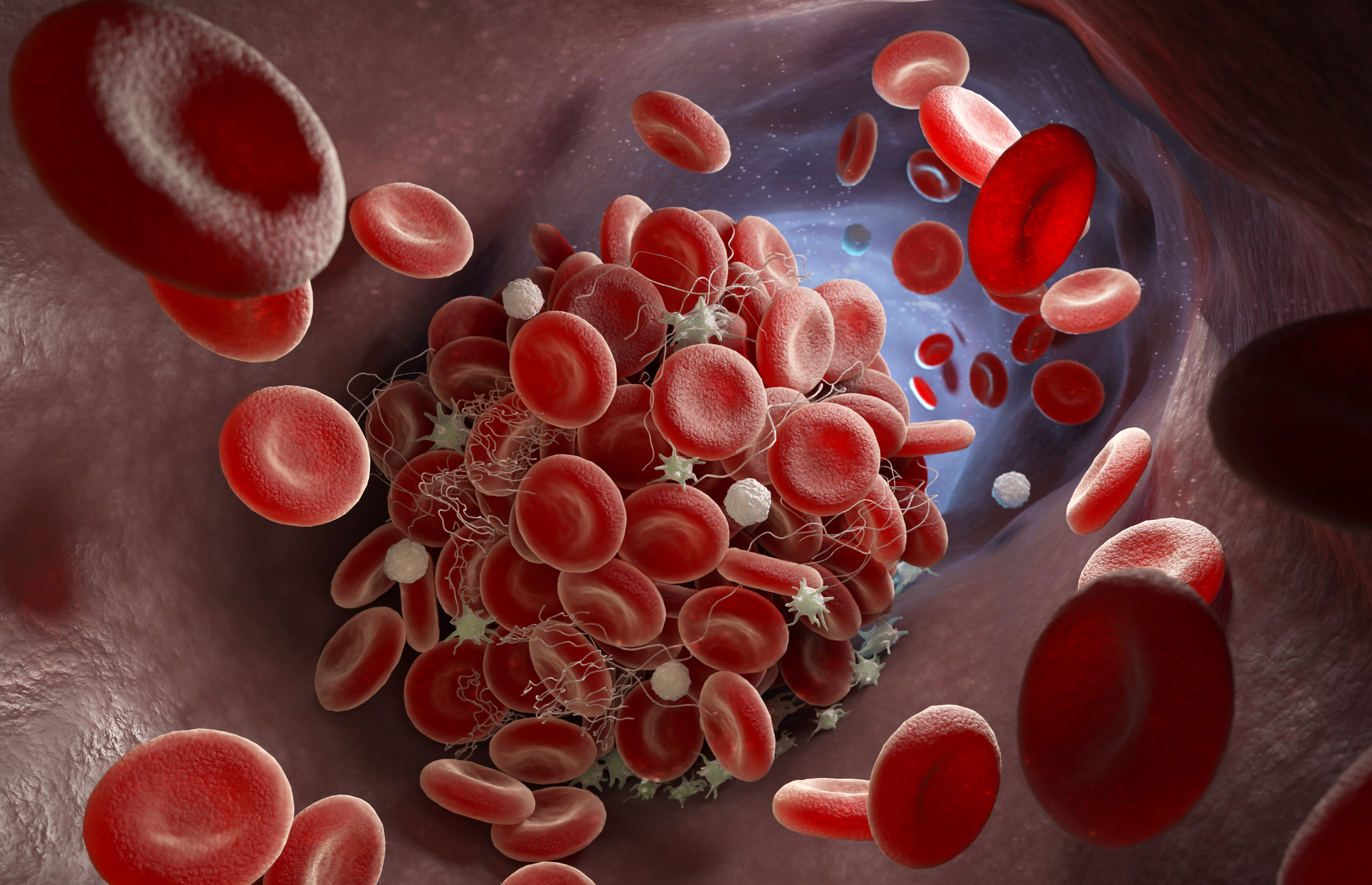
New tool to analyze blood platelets holds major medical potential
By Carol Clark
A novel technique to test platelet function within a person’s blood sample is faster, easier and more precise than methods currently in use, an experimental study shows.Thursday, October 19, 2023
Math trio makes new points about size of the smallest triangle
Friday, September 8, 2023
NIH funds Emory center to advance cellular mechanics
By Carol Clark
The National Institutes of Health (NIH) awarded Emory University $5.6 million to establish a national center to advance pioneering technology for cellular mechanics. The center is directed by Khalid Salaita, Emory professor of chemistry, whose lab developed the first sensors for detecting cell-receptor forces at the molecular level.
“We’ve been working on our molecular-force probes for more than a decade,” Salaita says. “We’ve demonstrated that these probes can be used to visualize, measure and map cellular forces down to the level of piconewtons. The center allows us to get this technology into the hands of end users — researchers in the biomedical sciences.”
The Center for Molecular Mechanobiology encompasses labs from seven leading research institutions including: Children’s Hospital of Philadelphia, Dana-Farber Cancer Institute, Emory, Georgia Tech, Memorial Sloan Kettering, University of Utah and Vanderbilt University.
The center members will use the molecular-force probes to investigate the biomechanics of processes such as the clotting of blood cells, the response of immune cells to an infection and the migration of cancer cells. Better understanding these processes may lead to the development of new treatments and therapies for a range of diseases and disorders.
In addition to supplying the technology, the center will train researchers to use the molecular-force probes and help adapt the technology to answer specific biomedical research questions.
“Working directly with the research community will help us to further refine and optimize the technology,” Salaita says. “We envision that measuring cellular forces will soon become part of the standard repertoire of biochemical techniques that scientists use to study living systems.”
The center’s associate directors are Yonggan Ke (associate professor in the Wallace H. Coulter Department of Biomedical Engineering at Emory and Georgia Tech) and Alexa Mattheyses (associate professor in the Department of Cell Developmental and Integrative Biology at the University of Alabama).
The five-year award from the National Institute of General Medical Sciences is part of the NIH Biomedical Technology Optimization and Dissemination Centers program. The goal is to optimize and disseminate state-of-the-art, late-stage biomedical technologies.
The first detailed view of mechanical forces
The Salaita lab works at the intersection of chemistry, biology and the physical sciences. It uses the building blocks of nature — nucleic acids — to create synthetic micro motors and probes for investigating fundamental questions of biology.
The molecular-force probes, developed by the Salaita lab in 2011, provide the first detailed view of the mechanical forces on the surface of a cell. The technology can detect mechanical forces as fleeting as the blink of an eye and as faint as piconewtons — about one billionth the weight of a paperclip.
The probes are made from strands of synthetic DNA tagged with fluorescence so that they function like molecular beacons, shining when they sense force. The technique is noninvasive, does not modify the cell and can be done with a standard fluorescence microscope.
In 2014, the lab used the new method to demonstrate how adherent cells — the kind that form the architecture of all multicellular organisms — mechanically sense their environments, migrate and stick to things.
In 2016, the molecular-force probes provided the first direct evidence for the mechanical forces of T cells, the security guards of the immune system. The lab’s experiments on T cells drawn from mice showed how they use a kind of mechanical “handshake” to test whether a cell they encounter is a friend or a foe.
In 2017, the lab shined its molecular beacons on platelets, the cells in the blood whose job is to stop bleeding by sticking together to form clots and plug up a wound. That work revealed the key molecular forces on platelets that trigger the clotting process.
In 2020, the lab and its collaborators combined advances in optical imaging with the molecular-force probes to capture forces at a resolution of 25 nanometers — far shorter than the length of a light wave. “That resolution is akin to being on the moon and seeing the ripples caused by raindrops hitting the surface of a lake on the Earth,” Salaita said at the time.
Key technological goals
The Center for Molecular Mechanobiology will build on this foundational work of the Salaita lab. It will focus on three key technological development goals:
• Optimizing the highest-resolution technique of the molecular-force probes so that it can be applied to a range of research questions.
• Tagging cells based on their force level in order to use force as a marker to barcode cells and their receptors. The idea is to classify the mechanics of individual cells and then link these classifications to gene-expression levels to study the cause-and-effect relationships.
• Amplifying the molecular-force signals to better understand the role of even the weakest forces involved in cellular mechanics, including those involved in the immune response.
Researchers from throughout the country will come to the Center for Molecular Mechanobiology to receive hands-on training in the molecular-force probes and then return to their home labs to become ambassadors for the technology.
“We’ll be adding a whole other layer of information for researchers working on everything from designing vaccines to cancer immunotherapy agents,” Salaita says.
Decades ago, he points out, complicated techniques such as crystallography, PCR and mass spectrometry were not frequently used but have since become routine workhorses in the biomedical sciences.
“We are catalyzing the process of spreading our technology so that studying biomechanics also becomes common and routine in biology,” Salaita says. “Molecular forces are a missing piece to understanding the way biology works.”
Related:
‘Firefly’ imaging method makes cellular forces visible
Chemists reveal the force within you
T cells use ‘handshakes’ to sort friends from foes
New methods reveal the mechanics of blood clotting
@font-face
{font-family:"Cambria Math";
panose-1:2 4 5 3 5 4 6 3 2 4;
mso-font-charset:0;
mso-generic-font-family:roman;
mso-font-pitch:variable;
mso-font-signature:-536870145 1107305727 0 0 415 0;}@font-face
{font-family:Calibri;
panose-1:2 15 5 2 2 2 4 3 2 4;
mso-font-charset:0;
mso-generic-font-family:swiss;
mso-font-pitch:variable;
mso-font-signature:-536859905 -1073732485 9 0 511 0;}p.MsoNormal, li.MsoNormal, div.MsoNormal
{mso-style-unhide:no;
mso-style-qformat:yes;
mso-style-parent:"";
margin:0in;
mso-pagination:widow-orphan;
font-size:12.0pt;
font-family:"Calibri",sans-serif;
mso-ascii-font-family:Calibri;
mso-ascii-theme-font:minor-latin;
mso-fareast-font-family:Calibri;
mso-fareast-theme-font:minor-latin;
mso-hansi-font-family:Calibri;
mso-hansi-theme-font:minor-latin;
mso-bidi-font-family:"Times New Roman";
mso-bidi-theme-font:minor-bidi;}a:link, span.MsoHyperlink
{mso-style-priority:99;
color:#0563C1;
mso-themecolor:hyperlink;
text-decoration:underline;
text-underline:single;}a:visited, span.MsoHyperlinkFollowed
{mso-style-noshow:yes;
mso-style-priority:99;
color:#954F72;
mso-themecolor:followedhyperlink;
text-decoration:underline;
text-underline:single;}.MsoChpDefault
{mso-style-type:export-only;
mso-default-props:yes;
font-family:"Calibri",sans-serif;
mso-ascii-font-family:Calibri;
mso-ascii-theme-font:minor-latin;
mso-fareast-font-family:Calibri;
mso-fareast-theme-font:minor-latin;
mso-hansi-font-family:Calibri;
mso-hansi-theme-font:minor-latin;
mso-bidi-font-family:"Times New Roman";
mso-bidi-theme-font:minor-bidi;
mso-font-kerning:0pt;
mso-ligatures:none;}div.WordSection1
{page:WordSection1;}
Monday, August 7, 2023
Physicists open new path to exotic form of superconductivity
By Carol Clark
Physicists have identified a mechanism for the formation of oscillating superconductivity known as pair-density waves. Physical Review Letters published the discovery, which provides new insight into an unconventional superconductive state seen in certain materials, including high-temperature superconductors.
“We discovered that structures known as Van Hove singularities can produce modulating, oscillating states of superconductivity,” says Luiz Santos, assistant professor of physics at Emory University and senior author of the study. “Our work provides a new theoretical framework for understanding the emergence of this behavior, a phenomenon that is not well understood.”
First author of the study is Pedro Castro, an Emory physics graduate student. Co-authors include Daniel Shaffer, a postdoctoral fellow in the Santos group, and Yi-Ming Wu from Stanford University.
The work was funded by the U.S. Department of Energy’s Office of Basic Energy Sciences.
The puzzle of superconductivity
Santos is a theorist who specializes in condensed matter physics. He studies the interactions of quantum materials — tiny things such as atoms, photons and electrons — that don’t behave according to the laws of classical physics.
Superconductivity, or the ability of certain materials to conduct electricity without energy loss when cooled to a super-low temperature, is one example of intriguing quantum behavior. The phenomenon was discovered in 1911 when Dutch physicist Heike Kamerlingh Onnes showed that mercury lost its electrical resistance when cooled to 4 Kelvin or minus 371 degrees Fahrenheit. That’s about the temperature of Uranus, the coldest planet in the solar system.
It took scientists until 1957 to come up with an explanation for how and why superconductivity occurs. At normal temperatures, electrons roam more or less independently. They bump into other particles, causing them to shift speed and direction and dissipate energy. At low temperatures, however, electrons can organize into a new state of matter.
“They form pairs that are bound together into a collective state that behaves like a single entity,” Santos explains. “You can think of them like soldiers in an army. If they are moving in isolation they are easier to deflect. But when they are marching together in lockstep it’s much harder to destabilize them. This collective state carries current in a robust way.”
A holy grail of physics
Superconductivity holds huge potential. In theory, it could allow for electric current to move through wires without heating them up, or losing energy. These wires could then carry far more electricity, far more efficiently.
“One of the holy grails of physics is room-temperature superconductivity that is practical enough for everyday-living applications,” Santos says. “That breakthrough could change the shape of civilization.”
Many physicists and engineers are working on this frontline to revolutionize how electricity gets transferred.
Meanwhile, superconductivity has already found applications. Superconducting coils power electromagnets used in magnetic resonance imaging (MRI) machines for medical diagnostics. A handful of magnetic levitation trains are now operating in the world, built on superconducting magnets that are 10 times stronger than ordinary electromagnets. The magnets repel each other when the matching poles face each other, generating a magnetic field capable of levitating and propelling a train.
The Large Hadron Collider, a particle accelerator that scientists are using to research the fundamental structure of the universe, is another example of technology that runs through superconductivity.
Superconductivity continues to be discovered in more materials, including many that are superconductive at higher temperatures.
An accidental discovery
One focus of Santos’ research is how interactions between electrons can lead to forms of superconductivity that cannot be explained by the 1957 description of superconductivity. An example of this so-called exotic phenomenon is oscillating superconductivity, when the paired electrons dance in waves, changing amplitude.
In an unrelated project, Santos asked Castro to investigate specific properties of Van Hove singularities, structures where many electronic states become close in energy. Castro’s project revealed that the singularities appeared to have the right kind of physics to seed oscillating superconductivity.
That sparked Santos and his collaborators to delve deeper. They uncovered a mechanism that would allow these dancing-wave states of superconductivity to arise from Van Hove singularities.
“As theoretical physicists, we want to be able to predict and classify behavior to understand how nature works,” Santos says. “Then we can start to ask questions with technological relevance.”
Some high-temperature superconductors — which function at temperatures about three times as cold as a household freezer — have this dancing-wave behavior.
The discovery of how this behavior can emerge from Van Hove singularities provides a foundation for experimentalists to explore the realm of possibilities it presents.
“I doubt that Kamerlingh Onnes was thinking about levitation or particle accelerators when he discovered superconductivity,” Santos says. “But everything we learn about the world has potential applications.”
Related:
Wednesday, July 26, 2023
Merck Prize boosts work on air sensor for pandemic pathogens
Merck KGaA, Darmstadt, Germany, awarded its 2023 Future Insight Prize to Khalid Salaita, professor of chemistry at Emory University. The award comes with $540,000 to fund the next phase of research into an air sensor that can continuously monitor indoor spaces for pathogens that can cause pandemics.
“I’m extremely thankful to receive the Future Insight Prize as this enables us to continue our path toward an early-warning system for emerging threats,” Salaita says. “Our research sets the stage for fully automated detection of airborne pathogens without human intervention or sample processing.”
The Merck Future Insight Prize recognizes groundbreaking ideas to solve some of the world’s most pressing challenges in health, nutrition and energy.
The Salaita lab’s sensor, a rolling micro-motor called “Rolosense,” holds the potential to help mitigate, or even prevent, a pandemic.
Tuesday, July 18, 2023
Biophysicists reveal how three proteins interact to fine-tune cellular movement
By Carol Clark
A single human cell teems with as many 100,000 different proteins. Actin is one of the most abundant and essential of them all. This protein forms into filaments that help make up the skeleton of cells, giving them shape. And as the actin filaments elongate, they work like muscles, pushing against the inner membrane of a cell to move it forward.
Three other proteins are known to drive the activities of actin. One class of protein assembles individual actin molecules into actin filaments, another causes the filaments to stop growing and a third disassembles filaments.
Biophysicists at Emory University, however, have discovered an even more complex and nuanced view of how these three proteins together influence actin dynamics. Nature Communications published the findings, showing how these proteins sometimes shift from solo or duet acts to perform as a trio, allowing them to fine-tune the activity of actin filaments.
The discovery opens another window onto the dynamics of cellular movement, which is key to processes ranging from stem-cell differentiation and wound healing to the development of diseases such as cancer.
“We found that while these three proteins do one thing when working on their own, they do a completely different thing when the other two proteins join them,” says Shashank Shekhar, Emory assistant professor of physics and cell biology, and senior author of the study. “It gets really complex, very fast.”
“No one had looked at all of these proteins interacting at once on actin,” adds Heidi Ulrichs, co-first author of the study and an Emory PhD candidate in biochemistry, cell and developmental biology. “Our paper is the first report of all three of them occupying the same barbed end of an actin filament.”
Ulrichs worked closely on the project with Ignas Gaska, a postdoctoral fellow in the Shekkhar lab who is co-first author of the paper.
Building on previous research
Research into how proteins act individually on actin is relatively well-characterized.
A polymerase protein, such as formin, drives elongation of actin. Formin positions itself at the end of an actin filament, grabs onto free-floating actin molecules and stacks them up one by one to keep growing the end.
Depolymerase proteins, such as twinfilin, are another class of proteins that influence actin. Twinfilin works like a lint roller, binding to the end of a filament and peeling away one molecule at a time. Twinfilin can repeat the process to disassemble the actin filament entirely.
Proteins known as cappers can stop the elongation and disassembling of the filaments. A capper attaches to the end of an actin filament and covers it like a hat, blocking activity by the other proteins.
This knowledge was built up by isolating one protein at a time to study how it influences actin. More recent studies have also shown simultaneous interactions between twinfilin and capping proteins.
A new approach using advanced technology
For the current study, the researchers wanted to explore whether formin, twinfilin and the capping protein could all three act simultaneously on actin.
“An actin filament end is really tiny, just five nanometers across,” Shekhar explains. “One thought was that there just isn’t enough real estate available for three proteins to work on a single actin filament at once.”
The Shekhar lab is one of only a handful in the world using the highly specialized technique of microfluidics-assisted total internal reflection fluorescence microscopy (mf-TIRF) to study how the actin cytoskeleton remodels itself.
Cells are packed with thousands of proteins moving around, performing different functions, making it impossible to track all of them. Researchers must isolate the proteins of interest and study them outside of a cellular system, by introducing them to a microfluidic system on a microscope slide.
The mf-TIRF technology allows the Shekhar Lab to attach fluorescent orbs to single protein molecules so that researchers can better observe what these molecules are doing through a microscope.
In experiments, the researchers tagged molecules of actin, formin, twinfilin and the capping protein with four different colors that emitted fluorescent light. They then introduced actin to the microfluidic system and added the other proteins one at a time.
Establishing a new paradigm
The results startled them.
When twinfilin, the protein that breaks apart an actin filament, was added in the presence of both formin and the capping protein, twinfilin actually worked to speed up the process of filament elongation.
“That’s counterintuitive, which is cool,” Ulrichs says. “Doing science you get surprised all the time.”
Twinfilin alone could not join formin on the end of the actin filament. However, when the capping protein was also present, all three could simultaneously work together on the tiny surface of the actin filament.
Shekhar compares the effects of all three proteins working together to a knob that allows for more precise control of a process.
“Our findings establish a new paradigm in which the three proteins work in concert to fine-tune how fast or slowly actin filaments are formed,” he says. The dynamics of how the three proteins interact with actin is fundamental to teasing apart the complex mysteries of how cells function normally and what happens when something goes wrong.
“We’re building up knowledge, step by step, study by study, on the dynamics of what’s happening inside of a cell,” Ulrichs says.
Related:
Wednesday, March 22, 2023
As the worm turns: New twists in behavioral association theories
By Carol Clark
Physicists have developed a dynamical model of animal behavior that may explain some mysteries surrounding associative learning going back to Pavlov’s dogs. The Proceedings of the National Academy of Sciences (PNAS) published the findings, based on experiments on a common laboratory organism, the roundworm C. elegans.
“We showed how learned associations are not mediated by just the strength of an association, but by multiple, nearly independent pathways — at least in the worms,” says Ilya Nemenman, an Emory professor of physics and biology whose lab led the theoretical analyses for the paper. “We expect that similar results will hold for larger animals as well, including maybe in humans.”
“Our model is dynamical and multi-dimensional,” adds William Ryu, an associate professor of physics at the Donnelly Centre at the University of Toronto, whose lab led the experimental work. “It explains why this example of associative learning is not as simple as forming a single positive memory. Instead, it’s a continuous interplay between positive and negative associations that are happening at the same time.”
First author of the paper is Ahmed Roman, who worked on the project as an Emory graduate student and is now a postdoctoral fellow at the Broad Institute. Konstaintine Palanski, a former graduate student at the University of Toronto, is also an author.
The conditioned reflex
More than 100 years ago, Ivan Pavlov discovered the “conditioned reflex” in animals through his experiments on dogs. For example, after a dog was trained to associate a sound with the subsequent arrival of food, the dog would start to salivate when it heard the sound, even before the food appeared.
About 70 years later, psychologists built on Pavlov’s insights to develop the Rescorla-Wagner model of classical conditioning. This mathematical model describes conditioned associations by their time-dependent strength. That strength increases when the conditioned stimulus (in Pavlov dog’s case the sound) can be used by the animal to decrease the surprise in the arrival of the unconditioned response (the food).
Such insights helped set the stage for modern theories of reinforcement learning in animals, which in turn enabled reinforcement learning algorithms in artificial intelligence systems. But many mysteries remain, including some related to Pavlov’s original experiments.
After Pavlov trained dogs to associate the sound of a bell with food he would then repeatedly expose them to the bell without food. During the first few trials without food, the dogs continued to salivate when the bell rang. If the trials continued long enough, the dogs “unlearned” and stopped salivating in response to the bell. The association was said to be “extinguished.”
Pavlov discovered, however, that if he waited a while and then retested the dogs, they would once again salivate in response to the bell, even if no food was present. Neither Pavlov nor more recent associative-learning theories could accurately explain or mathematically model this spontaneous recovery of an extinguished association.
Teasing out the puzzle
Researchers have explored such mysteries through experiments with C. elegans. The one-millimeter roundworm only has about 1,000 cells and 300 of them are neurons. That simplicity provides scientists with a simple system to test how the animal learns. At the same time, C. elegans’ neural circuitry is just complicated enough to connect some of the insights gained from studying its behavior to more complex systems.
Earlier experiments have established that C. elegans can be trained to prefer a cooler or warmer temperature by conditioning it at a certain temperature with food. In a typical experiment, the worms are placed in a petri dish with a gradient of temperatures but no food. Those trained to prefer a cooler temperature will move to the cooler side of the dish, while the worms trained to prefer a warmer temperature go to the warmer side.
But what exactly do these result mean? Some believe that the worms crawl toward a particular temperature in expectation of food. Others argue that the worms simply become habituated to that temperature, so they prefer to hang out there even without a food reward.
The puzzle could not be resolved due to a major limitation of many of these experiments — the lengthy amount of time it takes for a worm to traverse a nine-centimeter petri dish in search of the preferred temperature.
Measuring how learning changes over time
Nemenman and Ryu sought to overcome this limitation. They wanted to develop a practical way to precisely measure the dynamics of learning, or how learning changes over time.
Ryu’s lab used a microfluidic device to shrink the experimental model of nine-centimeter petri dishes into four-millimeter droplets. The researchers could rapidly run experiments on hundreds of worms, each worm encased within its individual droplet.
“We could observe in real time how a worm moved across a linear gradient of temperatures,” Ryu says. “Instead of waiting for it to crawl for 30 minutes or an hour, we could much more quickly see which side of the droplet, the cold side or the warm side, that the worm preferred. And we could also follow how its preferences changed with time.”
Their experiments confirmed that if a worm is trained to associate food with a cooler temperature it will move to the cooler side of the droplet. Over time, however, with no food present, this memory preference seemingly decays.
“We found that suddenly the worms wanted to spend more time on the warm side of the droplet,” Ryu says. “That’s surprising because why would the worms develop a different preference and even avoidance of the temperature they had come to associate with food?”
Eventually the worm begins moving back and forth between the cooler and warmer temperatures. The researchers hypothesized that the worm does not simply forget the positive memory of food associated with cooler temperatures but instead starts to negatively associate the cooler side with no food. That spurs it to head for the warmer side. Then as more time passes, it begins to form a negative association of no food with the warmer temperature, which combined with the residual positive association to the cold, makes it migrate back to the cooler one.
“The worm is always learning, all the time,” Ryu explains. “There is an interplay between the drive of a positive association and a negative association that causes it to start oscillating between cold and warm.”
'It's like when you lose your keys'
Nemenman’s team developed theoretical equations to describe the interactions over time between the two independent variables — the positive, or excitatory, association that drives a worm toward one temperature and the negative, or inhibitory, association that drives it away from that temperature.
“The side that the worm gravitates toward depends on when exactly you take the measurements,” Nemenman explains. “It’s like when you lose your keys you may check the desk where you usually keep them first. If you don’t see them there right away, you run around different places looking for them. If you still don’t find them, you go back to the original desk figuring you just didn’t look hard enough.”
The researchers repeated the experiments under different conditions. They trained the worms at different starting temperatures and starved them for different durations before testing their temperature preference, and the worms’ behaviors were correctly predicted by the equations.
They also tested their hypothesis by genetically modifying the worms, knocking out the insulin-like signaling pathway known to serve as a negative association pathway.
“We perturbed the biology in specific ways and when we ran the experiments, the worm’s behavior changed as predicted by our theoretical model,” Nemenman says. “That gives us more confidence that the model reflects the underlying biology of learning, at least in C. elegans.”
The researchers hope that others will test their model in studies of larger animals across species.
“Our model provides an alternative quantitative model of learning that is multi-dimensional,” Ryu says. “It explains results that are difficult, or in some cases impossible, for other theories of classical conditioning to explain.”
Related:
Physicists develop theoretical model for neural activity of mouse brain
Machine learning used to understand and predict dynamics of worm behavior
Friday, January 27, 2023
Emory physicists to study airborne microbes, funded by $1.2 million Keck Award
By Carol Clark
Microbes have an incredible ability to thrive in different environments. Extensive research has shown the vital roles that these invisible organisms play in the ecosystems of marine and soil environments. But the atmosphere is another important habitat.
Growing evidence shows that microbes in the Earth’s atmosphere can affect rainfall, land fertilization and food production. Little is known, however, about how these tiny life forms adapt to living in air and the even broader role that airborne microbes may play in the planet’s ecosystem.
The W.M. Keck Foundation awarded Emory University physicists Justin Burton and Minsu Kim $1.2 million to explore these mysteries. In a collaboration with the University of Oregon, the researchers will use the funds to pioneer new methods for mechanistic studies of the physiology, metabolism, ecology and evolution of airborne microbes.
The Emory project will create new tools to conduct never-before-done studies of how microbes adapt to living in air. “We’ve developed a prototype acoustic levitation system that opens the door for air-culturing microbes in a well-controlled laboratory environment for the first time,” says Burton, associate professor of physics.
The Burton lab specializes in studying the fluid dynamics of natural phenomena, from the molecular to the geographical scale.
The Kim lab specializes in using advanced biophysical techniques to characterize microbes from the molecular to cellular level.
“We are helping to take the field of air biology into a new era,” says Kim, associate professor of physics. “Most research into microbe ecology has focused on microbes from marine and soil environments. We’re expanding the possibilities for investigating atmospheric microbes.”
The Emory researchers will collaborate with additional principal investigators on the Keck Award project: Earth scientist Joshua Méndez Harper, a former postdoctoral fellow in the Burton lab now at the University of Oregon, and Josef Dufek, professor of Earth sciences at the University of Oregon.
Wind-blown dust circulates globally, emitted by volcanic eruptions, wildfires and dust storms. These airborne particles can carry minerals and other nutrients across long distances. Desert dust from the Sahara, for example, is an important source of phosphorous for the Amazon rain forest. It is also well-known that microbes — including viruses, fungi and bacteria — hitchhike on atmospheric dust.
Perhaps the most striking example of how atmospheric microbes can impact the environment is the so-called rain-making bacterium Pseudomonas syringae that has been isolated from clouds. Research shows that this bacterium may play a role in the precipitation cycle by producing an enzyme that catalyzes ice formation.
Key impediments to further studying such atmospheric phenomena are sampling and culturing microbes in the air. The Keck-funded project will develop an airborne-culturing method using acoustic levitators, each about the size of a microwave oven, that simulate atmospheric conditions. The levitators will work by creating standing waves of sound, just like a musical instrument, but at a frequency well outside of the range of human hearing. These high-intensity sound waves create high pressure that is capable of suspending particles in air that are as dense as copper.
Initially, the project will focus on studying bacteria, believed to make up more than 50% of atmospheric microbes. The controlled, laboratory conditions of the levitators will allow the researchers to home in on how different species of bacteria adapt to living in the harsh conditions found in the atmosphere, such as extreme temperature shifts, high humidity and solar radiation.
The bacterial species studied will include some that the researchers collect from the atmosphere through balloon experiments. Team members at the University of Oregon will release a series of small, ultralight balloons. They will float on easterly winds across the country at altitudes between five and six kilometers, which is comparable to those of Saharan dust clouds and wildfire plumes. Each balloon will house sterile titanium booms affixed with tiny sponges to trap microbes within their pores.
Balloon positions and sensor data — including temperature, pressure, humidity and dust concentration — will be transmitted continuously using amateur radio bands. As the balloons fly over Georgia, descent will be controlled using parachutes. GPS beacons will be enabled upon landing, allowing the researchers to pinpoint and recover the balloons.
The methods developed in the lab, and the resulting data, will be open source. Scientists around the world can build on the work and continue to push the boundaries in the field of air biology.
“The award from the Keck Foundation is giving us the freedom to strike out in bold new scientific directions,” Burton says.
Based in Los Angeles, the W.M. Keck Foundation was established in 1954 by the late W.M. Keck, founder of the Superior Oil Company. The foundation’s grant making is focused primarily on pioneering efforts in the areas of medical research and science and engineering. The foundation also supports undergraduate education and maintains a Southern California Grant Program that provides support for the Lost Angeles community, with a special emphasis on children and youth. For more information, visit www.wmkeck.org.
Related:
Soil quality critical to help some U.S. crops weather heat stress from climate change
Chemists crack complete quantum nature of water
Chemists map cascade of reactions for producing atmosphere's 'detergent'
Friday, September 9, 2022
Physicist seeks ultimate formula for fun
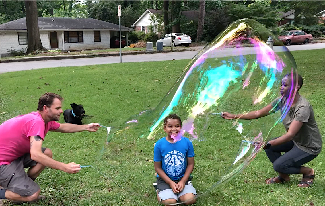
Justin and Afeira Burton encase their son Jonah in a giant bubble while their dog Boba Fetch looks on.
By day, Justin Burton is an Emory associate professor of physics, conducing high-level research on fluid dynamics and granular materials. Evenings and weekends, however, he turns into a comic-book version of a scientist. Not a mad scientist, tough. More like a glad scientist.
Read more about his alter ego, Dr. Bubbles, here.
Related:
Tuesday, August 23, 2022
Chronic COVID infections source of variants of concern, study shows
An electron microscopic image shows an isolate from the first U.S. case of COVID-19, caused by SARS-CoV-2. The virus has since evolved into five variants of concern. "If we want to stay a step ahead of this virus, we need to be more actively identifying and surveilling people with chronic infections," says Emory graduate Mahan Ghafari, first author of a new study on how the virus evolves. (CDC)
By Carol Clark
The coronavirus variants of concern are emerging from chronic, long-term COVID infections in people who may be immune comprised and unable to clear the virus, a new study strongly suggests. Frontiers in Virology published the findings by scientists at Emory University and the University of Oxford.
“Rather than evolving from transmission chains of acute COVID infections in hundreds of millions of people, our results show that the variants of concern come from rare cases when someone may have an active infection for months,” says Daniel Weissman, a corresponding author and Emory professor of biology and physics focused on quantitative evolutionary theory.
“A key take-home message is that it is important to find these individuals who are chronically infected and provide support for them to recover,” adds Mahan Ghafari, first author of the paper and a postdoctoral researcher at the University of Oxford. “In many cases they may be asymptomatic and not even realize that they are infected with COVID although they are actively shedding the virus.”
Ghafari graduated from Emory in 2018 with a masters in physics.
Additional authors of the paper include Aris Katzourakis, a professor of evolution and genomics at the University of Oxford; Qihan Liu, an Emory graduate student in physics; and Emory undergraduate Arushi Dhillon.
Random mutations
Viruses like SARS-CoV-2 continuously evolve due to occasional mutations in the genetic code that may occur when they replicate. “When a virus copies itself, it doesn’t always make perfect copies,” Weissman explains.
Usually, such random mutations do not benefit the virus or raise the concerns of scientists monitoring these changes. Occasionally, however, the mutations result in a variant of the virus that may make it more transmissible, more difficult to detect and treat, and even more lethal.
The World Health Organization defines a SARS-CoV-2 variant of concern as one that is more likely to cause infections even in those who are vaccinated or in those who were previously infected.
“During the first few months of the pandemic, it didn’t look like the coronavirus was going to adapt into a variant of concern,” Weissman says. “But then, boom, boom, boom! Not only did the coronavirus evolve into VOCs, it did it three times in quick succession in late 2020.”
The WHO dubbed these first three variants of concern alpha, beta and gamma.
Mysteries surrounding VOCs
Why had all three of these VOCs emerged at roughly the same time and apparently in three far-flung areas of the world?
Another mystery was why large clusters of mutations occurred in the VOCs. “A key element that distinguished these VOCs from other lineages of virus that were circulating is that each of them has a vastly elevated number of mutations,” Ghafari notes. “That’s a major distinction point in evolutionary terms.”
At least some of the mutations from the VOC had been detected in chronic cases of COVID, leading to the hypothesis that these long-term cases may be the source of the VOCs. The other main theory was that VOCs were emerging from sustained transmission of acute infections in areas of the world with poor genomic surveillance of the virus.
Ghafari, Weissman and their collaborators were among the first teams to methodically test these theories surrounding the emergence of the alpha, beta and gamma VOCs.
The researchers built a mechanistic, theoretical model to study the problem, using existing data and software they developed.
The resulting model rules out the theory that the VOCs emerged from sustained transmission of acute infections and fully supports the theory that each variant evolved within a single individual with a chronic infection.
The model shows how multiple mutations were needed, each of which may have been either neutral or slightly advantageous to viral fitness. In this way, a variant eventually acquires a constellation of mutations that allow it to become more transmissible.
Model explains delta and omicron emergence
Although the current paper drew from data for the alpha, beta and gamma variants, the resulting theoretical model also explains the later independent emergence of the delta and omicron VOCs. Delta emerged in India in late 2020, rapidly sweeping through that country and spreading around the world. Delta subsided after omicron, which is not a descendent of the delta variant, emerged in South Africa in late 2021. Omicron quickly became the dominant global VOC.
The researchers have made their model and software publicly available for others to study the evolution of SARS-CoV-2 variants.
“Ideally, we’d like to eventually be able to quantify the timing at which new variants might emerge in the future,” Weissman says. “That has huge implications from a public health perspective.”
Studies have shown that some immune-compromised people, such as those taking medication for other chronic disorders, have carried active COVID infections for a year or even longer. It’s critical to identify these people, the researchers stress, not only to help them get treated for COVID, but also to conduct genomic surveillance of the SARS-CoV-2 viruses that they carry.
“Who knows what variant could be boiling up next from a chronically infected individual?” Ghafari says. “Our study shows that from an evolutionary point of view, we can expect something completely different from the previous VOCs. If we want to stay a step ahead of this virus, we need to be more actively identifying and surveilling people with chronic infections.”
The work was funded by the National Science Foundation, the Simons Foundation, the Sloan Foundation, the Biotechnology and Biological Science Research Council and the European Research Council.
Related:
Viral sequencing can reveal how SARS-CoV-2 spreads and evolves
Emory physicist Daniel Weissman awarded 2021 Sloan Research Fellowship
Monday, July 11, 2022
Chemists crack complete quantum nature of water
Chemists have produced the first full quantum mechanical model of water — one of the key ingredients of life. The Journal of Physical Chemistry Letters published the breakthrough, which used machine learning to develop a model that gives a detailed, accurate description for how large groups of water molecules interact with one another.
“We believe we have found the missing piece to a complete, microscopic understanding of water,” says Joel Bowman, professor of theoretical chemistry at Emory University and senior author of the study. “It appears that we now have all that we need to know to describe water molecules under any conditions, including ice, liquid or vapor over a range of temperature and pressure.”
The researchers developed free, open-source software for the model, which they dubbed “q-AQUA.” The q-AQUA software provides a universal tool for studying water.
“We anticipate researchers using it for everything from predicting whether an exoplanet may have water to deepening our understanding of the role of water in cellular function,” Bowman says.
Read more about the discovery here.
Related:
Chemists map cascade of reactions for producing atmosphere's 'detergent'